Phage , the virus that cures

Page 11 - Seeking answers? Join the AnandTech community: where nearly half-a-million members share solutions and discuss the latest tech.
May 11, 2008
19,469
1,159
126
Bacteria that can transport electrons over "vast" distances.
It seems i am experiencing the vast range of symptoms of the current flu epidemic.
For now, this is all the commenting i can provide for i feel largely depleted of energy.

http://www.nature.com/nature/journal/v491/n7423/fig_tab/nature11586_F1.html

nature11586-f1.2.jpg


http://en.wikipedia.org/wiki/Desulfobulbaceae
The discovery of filamentous Desulfobulbaceae in 2012 elucidates the cause of the small electrical currents in the top layer of sediment on large portions of the ocean floor. The currents were first measured in 2010. Thousands of these currently unnamed Desulfobulbus cells are arranged in fibrous microorganisms up to a centimeter in length. They transport electrons from the sediment that is rich in hydrogen sulfide up to the oxygen-rich sediment that is in contact with the water.

http://www.wired.com/wiredscience/2012/10/bacteria-electric-wires


The world's deep seafloors are dark and airless places, but vast swaths may pulse gently with energy conducted through a type of newly discovered bacteria that forms living electrical cables.
The bacteria were first detected in 2010 by researchers perplexed at chemical fluctuations in sediments from the bottom of Aarhus Bay in Denmark. Almost instantaneously linking changing oxygen levels in water with reactions in mud nearly an inch below, the fluctuations occurred too fast to be explained by chemistry.
Only an electrical signal made sense -- but no known bacteria could transmit electricity across such comparatively vast distances. Were bacteria the size of humans, the signals would be making a journey 12 miles long.
Now the mysterious bacteria have been identified. They belong to a microbial family called Desulfobulbaceae, though they share just 92 percent of their genes with any previously known member of that family. They deserve to be considered a new genus, the study of which could open a new scientific frontier for understanding the interface of biology, geology and chemistry across the undersea world.
The bacteria are described Oct. 24 in Nature by researchers led by microbiologists Christian Pfeffer, Nils Risgaard-Petersen and Lars Peter Nielsen of Aarhus University. On the following pages, Wired takes a look at these marvelous microbes.
Seen through an electron microscope, the Desulfobulbaceae -- the researchers haven't yet given them a genus or species name -- appear in blue. They link end-to-end, forming filaments nearly an inch in length.

http://www.wired.com/wiredscience/2010/02/electric-ocean-bacteria/
According to findings that could have been pulled from a deep-sea sequel to Avatar, bacteria appear to conduct electrical currents across the ocean floor, driving linked chemical reactions at relatively vast distances.
Noticed only when reseachers happened to test sediment leftovers from another experiment, the phenomenon may add a new mechanism to Earth’s biogeochemistry.
“The cycling of elements and life at the bottom of the sea, and in soil, and anywhere else you’re short of oxygen — this could help us understand those processes,” said microbiologist Lars Peter Nielsen of Denmark’s Aarhus University, co-author of the study, published Feb. 24 in Nature.
The original focus of Nielsen’s team wasn’t seafloor conductivity, but an especially interesting species of sulfur bacteria found on the floor of Aarhus Bay. To help quantify their chemical activity, the researchers kept a few beakers of seawater and sulfur bacteria-free sediment for comparison.
After those experiments ended, the beakers were almost forgotten. Then, a few weeks later, the researchers noticed strange patterns of activity. Changing oxygen levels in water above the top sediment layer were almost immediately followed by chemical fluctuations several layers down. The distance was so great, and the response time so quick, that usual methods of chemical transport — molecular diffusion, or a slow drift from high to low concentration — couldn’t explain it.
At first, the researchers were stumped. Then they realized the process made sense if bacteria in the top and bottom layers were linked. Anything that affected oxygen-processing bacteria up top would also affect the sulfide-eating microbes below. It would explain the apparent connection; and an electrical linkage would explain the speed. It would also boggle the mind.
“Such hypotheses would at one time have been considered heretical,” wrote Kenneth Nealson, a University of Southern California microbiologist, in an accompanying commentary in Nature. A half-inch gap “doesn’t seem like much of a distance. But to a bacterium it amounts to 10,000 body lengths, equivalent to about 20 kilometers (12 miles) in human terms.”
In recent years, however, scientists have found species of microbes with outer membranes covered by electron-transporting enzymes, or studded with conductive, micrometer-scale filaments. These are used in driving experimental microbial fuel cells, and are known to be found in the Aarhus Bay mud. Those sediments also contain trace amounts of pyrite, an electrically conductive mineral.
The top sediment layer also had a low concentration of hydrogen ions, something that could only be explained through an electrochemical reaction, with electrons conducted from a distance, said Nielsen.
Nealson called the findings “astonishing,” and said they “may be relevant to energy transfer and electron flow through many different environments.” They could eventually applied to bacteria-based schemes for bioremediation, carbon sequestration and energy production.
Asked if he’d seen the blockbuster movie Avatar, with its storyline involving electrochemically linked forests that stored the inhabitants’ souls in a planet-spanning biological computer, Nielsen said, “One of my colleagues saw this, and immediately sent me a message: ‘You’ve discovered the secret of Avatar! Go see it!’ The similarities are quite striking.”
He continued, “I don’t think there is much spirit in the networks we’ve seen here. It might be only about energy. But there are connections.”
 
May 11, 2008
19,469
1,159
126
I created this thread once long ago out of personal interest and to show to others that are interested as well... All the wonders of microscopic life and the impact that microscopic life has on us in every day life.
I never knew about the biologist Lynn Margulis who had already drawn the conclusion that becomes so apparent when reading this entire thread. How much microscopic life has an impact.
Something that is easily ignored. Life on this planet (and i am sure even in our solar system) can exist without dinosaurs or mammals. But life cannot exist without bacteria, viruses, fungi, single celled organisms.
I am waiting for the cover of the Madonna song "We are living in a material world". It will not be long before the cover "We are living in a bacterial world" will appear. :biggrin:


A wiki about the Late Lynn Margulis.
http://en.wikipedia.org/wiki/Lynn_Margulis
The percentage of the human genome that arose at a series of stages in evolution. 37% of human genes originated in bacteria.
animalsandbacteria1.jpg

Bacteria in an animal’s microbiota, such as those in the gut, in the mouth, and on the skin, communicate among themselves and exchange signals with the animal’s organ systems.
Some of the chemical signals are noted in this illustration. Credit: Margaret McFall-Ngai, et al. ©2013 PNAS
animalsandbacteria2.jpg

You can read the entire article here at phys.org.
http://phys.org/news/2013-02-bacterial-world-impacting-previously-thought.html#ajTabs

An excerpt :
Credit: Margaret McFall-Ngai, et al. ©2013 PNAS (Phys.org)—Throughout her career, the famous biologist Lynn Margulis (1938-2011) argued that the world of microorganisms has a much larger impact on the entire biosphere—the world of all living things—than scientists typically recognize. Now a team of scientists from universities around the world has collected and compiled the results of hundreds of studies, most from within the past decade, on animal-bacterial interactions, and have shown that Margulis was right. The combined results suggest that the evidence supporting Margulis' view has reached a tipping point, demanding that scientists reexamine some of the fundamental features of life through the lens of the complex, codependent relationships among bacteria and other very different life forms.
The project to review the current research on animal-bacterial interactions began when some scientists recognized the importance of bacteria in their own fields of study. For Michael Hadfield, Professor of Biology at the University of Hawaii at Manoa, the recognition grew over many years while studying the metamorphosis of marine animals. He found that certain bacteria influence marine larvae to settle to particular places on the sea floor, where they transform into juveniles and live out the rest of their lives. "Once we determined that specific biofilm bacteria provide an essential and unique ligand to stimulate the larvae of one globally distributed marine worm, our research naturally progressed to a study of the portion of the bacterial genome responsible for the signaling, and to other species, where we found the same genes involved," Hadfield told Phys.org. "Coming from different perspectives on the study of animal-bacterial interactions, and recognizing many more, Margaret McFall-Ngai [Professor of Medical Microbiology and Immunology at the University of Wisconsin, Madison] and I discussed the current situation extensively and then decided to attempt to draw together a significant number of experts on various approaches to the study of bacterial-animal interactions to draft a paper such as the one you have in hand. We proposed a 'catalysis meeting' on the subject to the National Science Foundation's National Evolutionary Synthesis Center (NESCent), which was funded, and the project took off." Bacteria surround us In many respects, it's easy to see the prominent role that bacteria play in the world. Bacteria were one of the first life forms to appear on Earth, about 3.8 billion years ago, and they will most likely survive long after humans are gone. In the current tree of life, they occupy one of the three main branches (the other two are Archaea and Eucarya, with animals belonging to the latter). Although bacteria are extremely diverse and live nearly everywhere on Earth, from the bottom of the ocean to the inside of our intestines, they have a few things in common. They are similar in size (a few micrometers), they are usually made of either a single cell or a few cells, and their cells don't have nuclei. Although scientists have known for many years that animals serve as a host for bacteria, which live especially in the gut/intestines, in the mouth, and on the skin, recent research has uncovered just how numerous these microbes are. Studies have shown that humans have about 10 times more bacterial cells in our bodies than we have human cells. (However, the total bacteria weigh less than half a pound because bacterial cells are much smaller than human cells.) While some of these bacteria simply live side-by-side with animals, not interacting much, some of them interact a lot. We often associate bacteria with disease-causing "germs" or pathogens, and bacteria are responsible for many diseases, such as tuberculosis, bubonic plague, and MRSA infections. But bacteria do many good things, too, and the recent research underlines the fact that animal life would not be the same without them. "The true number of bacterial species in the world is staggeringly huge, including bacteria now found circling the Earth in the most upper layers of our atmosphere and in the rocks deep below the sea floor," Hadfield said. "Then add all of those from all of the possible environments you can think of, from cesspools to hot springs, and all over on and in virtually every living organism. Therefore, the proportion of all bacterial species that is pathogenic to plants and animals is surely small. I suspect that the proportion that is beneficial/necessary to plants and animals is likewise small relative to the total number of bacteria present in the universe, and surely most bacteria, in this perspective, are 'neutral.' However, I am also convinced that the number of beneficial microbes, even very necessary microbes, is much, much greater than the number of pathogens." Animal origins and coevolution From our humble beginnings, bacteria may have played an important role by assisting in the origins of multicellular organisms (about 1-2 billion years ago) and in the origins of animals (about 700 million years ago). Researchers have recently discovered that one of the closest living relatives of multicellular animals, a single-celled choanoflagellate, responds to signals from one of its prey bacterium. These signals cause dividing choanoflagellate cells to retain connections, leading to the formation of well-coordinated colonies that may have become multicellular organisms. However, such questions of origin have been subjects of intense debate, and scientists have many hypotheses about how these life forms emerged. A bacterial role in these processes does not exclude other perspectives but adds an additional consideration.

After helping get animals started, bacteria also played an important role in helping them along their evolutionary path. While animal development is traditionally thought to be directed primarily by the animal's own genome in response to environmental factors, recent research has shown that animal development may be better thought of as an orchestration among the animal, the environment, and the coevolution of numerous microbial species. One example of this coevolution may have occurred when mammals evolved endothermy, or the ability to maintain a constant temperature of approximately 40 °C (100 °F) by metabolic means. This is also the temperature at which mammals' bacterial partners work at optimum efficiency, providing energy for the mammals and reducing their food requirement. This finding suggests that bacteria's preferred temperature may have placed a selection pressure on the evolution of genes associated with endothermy. Bacterial signaling Evidence for a deep-rooted alliance between animals and bacteria also emerges in both groups' genomes. Researchers estimate that about 37% of the 23,000 human genes have homologs with bacteria and Archaea, i.e., they are related to genes found in bacteria and Archaea that were derived from a common ancestor. Many of these homologous genes enable signaling between animals and bacteria, which suggests that they have been able to communicate and influence each other's development. One example is Hadfield and his group's discovery that bacterial signaling plays an essential role in inducing metamorphosis in some marine invertebrate larvae, where the bacteria produce cues associated with particular environmental factors. Other studies have found that bacterial signaling influences normal brain development in mammals, affects reproductive behavior in both vertebrates and invertebrates, and activates the immune system in tsetse flies. The olfactory chemicals that attract some animals (including humans) to their prospective mates are also produced by the animals' resident bacteria. Bacterial signaling is not only essential for development, it also helps animals maintain homeostasis, keeping us healthy and happy. As research has shown, bacteria in the gut can communicate with the brain through the central nervous system. Studies have found that mice without certain bacteria have defects in brain regions that control anxiety and depression-like behavior. Bacterial signaling also plays an essential role in guarding an animal's immune system. Disturbing these bacterial signaling pathways can lead to diseases such as diabetes, inflammatory bowel disease, and infections. Studies also suggest that many of the pathogens that cause disease in animals have "hijacked" these bacterial communication channels that originally evolved to maintain a balance between the animal and hundreds of beneficial bacterial species. Signaling also appears in the larger arena of ecosystems. For example, bacteria in flower nectar can change the chemical properties of the nectar, influencing the way pollinators interact with plants.

Human infants who are born vaginally have different gut bacteria than those delivered by Caesarean section, which may have long-lasting effects. And bacteria feeding on dead animals can repel animal scavengers—organisms 10,000 times their size—by producing noxious odors that signal the scavengers to stay away. In the gut In the earliest animals, gut bacteria played an important role in nutrition by helping animals digest their food, and may have influenced the development of other nearby organ systems, such as the respiratory and urogenital systems. Likewise, animal evolution likely drove the evolution of the bacteria, sometimes into highly specialized niches. For example, 90% of the bacterial species in termite guts are not found anywhere else. Such specialization also means that the extinction of every animal species results in the extinction of an unknown number of bacterial lineages that have evolved along with it. Scientists have also discovered that bacteria in the human gut adapts to changing diets. For example, most Americans have a gut microbiome that is optimized for digesting a high-fat, high-protein diet, while people in rural Amazonas, Venezuela, have gut microbes better suited for breaking down complex carbohydrates. Some people in Japan even have a gut bacterium that can digest seaweed. Researchers think the gut microbiome adapts in two ways: by adding or removing certain bacteria species, and by transferring the desired genes from one bacterium to another through horizontal gene transfer. Both host and bacteria benefit from this kind of symbiotic relationship, which researchers think is much more widespread than previously thought. The big picture Altogether, the recent studies have shown that animals and bacteria have histories that are deeply intertwined, and depend on each other for their own health and well-being as well as that of their environments. Although the researchers focused exclusively on animal-bacteria interactions, they expect that similar trends of codependency and symbiosis are universal among and between other groups, such as Archaea, fungi, plants, and animals. Once considered an exception, such intermingling is now becoming recognized as the rule—just as Margulis predicted many decades ago. Due to these symbiotic relationships, the scientists here propose that the very definitions of an organism, an environment, a population, and a genome have become blurred and should be reviewed. It may be, for instance, that animals are better viewed as host-microbe ecosystems than as individuals.

insect (1 mm) living in a forest canopy (10 m) illustrates the effects animal-bacterial interactions across multiple scales. Bacteria (1 micrometer) residing in the animal’s gut (0.1 mm) are essential to the insect’s nutrition, and insects often make up a majority of the animal biomass in forest canopies. Credit: Margaret McFall-Ngai, et al. ©2013 PNAS
animalsandbacteria3.jpg


In addition, the scientists predict that the recent findings on animal-bacteria interactions will likely require biologists to significantly alter their view of the fundamental nature of the entire biosphere. Along these lines, large-scale research projects such as the Human Microbiome Project and the Earth Microbiome Project are already underway to investigate the wide range of bacteria in the individual and global systems, and to see what happens when the bacteria are disturbed. In the end, the scientists hope that the results will promote more cross-disciplinary collaboration among scientists and engineers from different fields to explore the new microbial frontier. They argue that these discoveries should revolutionize the way that biology is taught from the high school level on up, by focusing more on the relationships between bacteria, their animal partners, and all other life forms. "It is hard to summarize a single 'most important conclusion,' other than the admonition to biologists studying animals, from behavior to physiology and ecology to molecular biology, that no matter what process you think you are studying, you must look for and consider a major role for bacteria," Hadfield said. "In many cases, this may require partnerships across traditional boundaries of research, meaning that zoologists must collaborate with microbiologists to advance their research, that molecular biologists must collaborate with whole-organism biologists, etc. We want badly for the message in 'Animals in a bacterial world,' to be a call for the necessary disappearance of the old boundaries between life science departments (e.g., Depts of Zoology, Botany, Microbiology, etc.) in universities, and societies (e.g., the American Society for Microbiology, etc.). We also want the message disseminated in college and university classes from introductory biology to advanced courses in the various topic areas of our paper." The results will profoundly change the way that the scientists of this collaboration continue with their own areas of research, Hadfield said. "Each of the authors of our paper conducts basic research in one or more areas of animal-bacterial interactions discussed in the paper, and each will continue to focus on her/his own speciality, I'm sure," he said. "However, I'm also certain that the interactions developed during the composition and writing of the paper (starting with our NESCent meeting in October 2011, when most of us met for the first time) will impact our own research and cause us to establish new collaborations with other laboratories. That has already occurred for me; I have a new collaboration with Dianne Newman's group at CalTech, an outstanding group of bacteriologists who are helping us do a much more in-depth investigation of the bacterial gene-products responsible for larval development."

More information: Margaret McFall-Ngai, et al. "Animals in a bacterial world, a new imperative for the life sciences." PNAS Early Edition. DOI: 10.1073/pnas.1218525110
Journal reference: Proceedings of the National Academy of Sciences
 
Last edited:
May 11, 2008
19,469
1,159
126
Bacteria produce their own set of tools to process a soluble form of uranium.
In reality, bacteria produce tiny nano-particles and use these nano particles to convert uranium from one form to another. Using this as part of their food cycle using redox reactions where electrons are "stripped" from one atom and passed on to another atom of a molecule.

I assume here, that when bacteria feed, they actually breakdown molecules by breaking apart the atoms that form covalent bonding by stripping electrons, weakening the atomic crystal.

Well, you can read the full story here :

http://phys.org/news/2013-03-unexpected-allies-bacteria-uranium-groundwater.html

(Phys.org) —Since 2009, SLAC scientist John Bargar has led a team using synchrotron-based X-ray techniques to study bacteria that help clean uranium from groundwater in a process called bioremediation. Their initial goal was to discover how the bacteria do it and determine the best way to help, but during the course of their research the team made an even more important discovery: Nature thinks bigger than that. The researchers discovered that bacteria don't necessarily go straight for the uranium, as was often thought to be the case. The bacteria make their own, even tinier allies – nanoparticles of a common mineral called iron sulfide. Then, working together, the bacteria and the iron sulfide grab molecules of a highly soluble form of uranium known as U(VI), or hexavalent uranium, and transform them into U(IV), a less-soluble form that's much less likely to spread through the water table. According to Barger, this newly discovered partnership may be the basis of a global geochemical process that forms deposits of uranium ore. And it's all done using one of the most basic types of chemical reactions known: oxidation and reduction, commonly known as "redox." Redox reactions can be thought of as the transfer of electrons from donor atoms to atoms that are hungry for electrons, and they are a primary source of chemical energy for both living and non-living processes. Photosynthesis involves redox reactions, as does cell respiration. Iron oxidizes to form rust; batteries depend on redox reactions to store and release energy. "Redox transitions are a very fundamental process," Bargar said. "It's the stuff of life. It's how you breathe." The study, published Monday in the Proceedings of the National Academy of Sciences, was conducted at the Old Rifle site on the Colorado River, a former uranium ore processing site in the town of Rifle, Colo. The aquifer at the site is contaminated with uranium and is the focus of bioremediation field studies conducted by a larger team of scientists at Lawrence Berkeley National Laboratory and funded by the Department of Energy's Office of Biological and Environmental Research. As part of their study, the LBNL team added acetate – essentially vinegar – to the aquifer in a series of injection wells to "feed the bugs," as Bargar put it, allowing acetate to flow throughout the aquifer around the wells. The SLAC team wanted to know what happened to the uranium during bioremediation. "We didn't want to study uranium only in the lab," Bargar said. "We wanted to understand uranium redox behavior in a living, breathing aquifer." They saw a bigger picture, including a molecular- to micron-scale view of what happened to other elements in the aquifer, such as sulfur and iron. During a series of redox reactions, the microbes dine on the acetate, and then pass extra electrons from their vinegar meal to – among other substances – naturally occurring sulfates. This liberates sulfur from the sulfates. A closer look at the soil, provided by X-ray microscopy images taken at the Stanford Synchrotron Radiation Lightsource and electron microscopy images recorded by collaborators at the Swiss Federal Institutes of Technology in Lausanne, Switzerland, revealed that the sulfur combined with iron in the soil to form iron sulfide nanoparticles, which did the actual work of transforming the uranium. At the same time, organic polymers produced by bacteria grabbed the transformed uranium and immobilized it. Discovering that bacteria work together with minerals to transform uranium was a surprise, said Bargar. Bargar said the discovery that a variety of processes immobilize the uranium sheds light on previous, seemingly conflicting observations, and also explains how the process can continue even when the uranium-munching bugs are in short supply. Better understanding Nature's methods for concentrating uranium could also lead to more efficient, environmentally friendly methods for uranium mining. But as a scientist, he appreciates the glimpse he's been given into Nature's abilities to multitask. "Originally we wanted to see what happened to uranium and how it could help bioremediation technology to be successful," he said. "But scientifically the results are much deeper than that." And since their original hypothesis focused on bacteria alone, it's a little humbling, too. "As is usual with science," said Bargar, "you learn your original ideas were a little naive, but finding out what's really going on is very exciting." Journal reference: Proceedings of the National Academy of Sciences Provided by SLAC National Accelerator Laboratory

http://en.wikipedia.org/wiki/Covalent_bond
 
Last edited:
May 11, 2008
19,469
1,159
126
Perhaps more proof how much a healthy diet and how this influences gut bacteria, seems to influence the proper functioning of the brain ?
I heard a few months ago on the radio a story about certain research, how the average IQ seems to become lower over time with humans. Perhaps it is diet related ? And what about diseases that are labeled as purely (psychological). This might give more insight and why there is an explosion of ADHD diagnosed people and also Asperger syndrome (autism spectrum)diagnosed people.

Imagine having stress. How much trouble you have concentrating or staying relaxed while having a lot of work or while being present in a social situation were you do not know what to do.
Now imagine that your body is experiencing severe stress because of your digestive system taking blow after blow because of an unhealthy and unbalanced gut fauna.
How much concentration and tolerance do you have left when you encounter situations that require all your attention... Knowing that some gut bacteria and some fungi can release toxins or waste products that seem to be similar to certain neurotransmitters (or even are just exactly neurotransmitters)or might just be plain neurotoxins... ^_^


http://medicalxpress.com/news/2013-05-gut-bacteria-diet-affects-brain.html

UCLA researchers now have the first evidence that bacteria ingested in food can affect brain function in humans. In an early proof-of-concept study of healthy women, they found that women who regularly consumed beneficial bacteria known as probiotics through yogurt showed altered brain function, both while in a resting state and in response to an emotion-recognition task.

The study, conducted by scientists with UCLA's Gail and Gerald Oppenheimer Family Center for Neurobiology of Stress and the Ahmanson–Lovelace Brain Mapping Center at UCLA, appears in the June edition of the peer-reviewed journal Gastroenterology.

The discovery that changing the bacterial environment, or microbiota, in the gut can affect the brain carries significant implications for future research that could point the way toward dietary or drug interventions to improve brain function, the researchers said.

"Many of us have a container of yogurt in our refrigerator that we may eat for enjoyment, for calcium or because we think it might help our health in other ways," said Dr. Kirsten Tillisch, an associate professor of medicine at UCLA's David Geffen School of Medicine and lead author of the study. "Our findings indicate that some of the contents of yogurt may actually change the way our brain responds to the environment. When we consider the implications of this work, the old sayings 'you are what you eat' and 'gut feelings' take on new meaning."

Researchers have known that the brain sends signals to the gut, which is why stress and other emotions can contribute to gastrointestinal symptoms. This study shows what has been suspected but until now had been proved only in animal studies: that signals travel the opposite way as well.

"Time and time again, we hear from patients that they never felt depressed or anxious until they started experiencing problems with their gut," Tillisch said. "Our study shows that the gut–brain connection is a two-way street."

The small study involved 36 women between the ages of 18 and 55. Researchers divided the women into three groups: one group ate a specific yogurt containing a mix of several probiotics—bacteria thought to have a positive effect on the intestines—twice a day for four weeks; another group consumed a dairy product that looked and tasted like the yogurt but contained no probiotics; and a third group ate no product at all.


Functional magnetic resonance imaging (fMRI) scans conducted both before and after the four-week study period looked at the women's brains in a state of rest and in response to an emotion-recognition task in which they viewed a series of pictures of people with angry or frightened faces and matched them to other faces showing the same emotions. This task, designed to measure the engagement of affective and cognitive brain regions in response to a visual stimulus, was chosen because previous research in animals had linked changes in gut flora to changes in affective behaviors.

The researchers found that, compared with the women who didn't consume the probiotic yogurt, those who did showed a decrease in activity in both the insula—which processes and integrates internal body sensations, like those form the gut—and the somatosensory cortex during the emotional reactivity task.

Further, in response to the task, these women had a decrease in the engagement of a widespread network in the brain that includes emotion-, cognition- and sensory-related areas. The women in the other two groups showed a stable or increased activity in this network.

During the resting brain scan, the women consuming probiotics showed greater connectivity between a key brainstem region known as the periaqueductal grey and cognition-associated areas of the prefrontal cortex. The women who ate no product at all, on the other hand, showed greater connectivity of the periaqueductal grey to emotion- and sensation-related regions, while the group consuming the non-probiotic dairy product showed results in between.

The researchers were surprised to find that the brain effects could be seen in many areas, including those involved in sensory processing and not merely those associated with emotion, Tillisch said.

The knowledge that signals are sent from the intestine to the brain and that they can be modulated by a dietary change is likely to lead to an expansion of research aimed at finding new strategies to prevent or treat digestive, mental and neurological disorders, said Dr. Emeran Mayer, a professor of medicine, physiology and psychiatry at the David Geffen School of Medicine at UCLA and the study's senior author.

"There are studies showing that what we eat can alter the composition and products of the gut flora—in particular, that people with high-vegetable, fiber-based diets have a different composition of their microbiota, or gut environment, than people who eat the more typical

Western diet that is high in fat and carbohydrates," Mayer said. "Now we know that this has an effect not only on the metabolism but also affects brain function."

The UCLA researchers are seeking to pinpoint particular chemicals produced by gut bacteria that may be triggering the signals to the brain. They also plan to study whether people with gastrointestinal symptoms such as bloating, abdominal pain and altered bowel movements have improvements in their digestive symptoms which correlate with changes in brain response.

Meanwhile, Mayer notes that other researchers are studying the potential benefits of certain probiotics in yogurts on mood symptoms such as anxiety. He said that other nutritional strategies may also be found to be beneficial.

By demonstrating the brain effects of probiotics, the study also raises the question of whether repeated courses of antibiotics can affect the brain, as some have speculated. Antibiotics are used extensively in neonatal intensive care units and in childhood respiratory tract infections, and such suppression of the normal microbiota may have longterm consequences on brain development.

Finally, as the complexity of the gut flora and its effect on the brain is better understood, researchers may find ways to manipulate the intestinal contents to treat chronic pain conditions or other brain related diseases, including, potentially, Parkinson's disease, Alzheimer's disease and autism.

Answers will be easier to come by in the near future as the declining cost of profiling a person's microbiota renders such tests more routine, Mayer said.

Explore further: Study: Probiotics reduce stress-induced intestinal flare-ups
 
Last edited:
May 11, 2008
19,469
1,159
126
Some parasites i mentioned in this thread, were able to hijack the host. Lucky for us humans, these are insects. However, some parasites uses mammals as well in their life cycle.

Dicrocoelium dendriticum, uses a snail and an ant to end up in cattle, the host.
When the ants are infected, these ants wonder around in the grass field and likely end up being consumed along with the grass by cattle. The big questions is of course, is Dicrocoelium dendriticum able to steer infected ants and infected snails in their movement ? It seems to be...

http://en.wikipedia.org/wiki/Dicrocoelium_dendriticum
 
May 11, 2008
19,469
1,159
126
Sigh... Some of the links in this thread are no longer functional. Lucky i copied the text.

Norovirus. As any person , i to get occasionally infected. And since it is winter, the noroviruses and adenoviruses are getting busy. And since i travel by use of public transport, my immunesystem is put to the test on a daily basis. Especially when it is crowded and there is bad air circulation or air conditioning.
Yesterday i was the unlucky subject to a norovirus infection(At least, i assume that) I do seem to have had all the symptoms. However, i read about adeno viruses that these can also cause gastroenteritis (infectious diarrhea).

http://en.wikipedia.org/wiki/Norovirus
Norovirus is a genus of genetically diverse single-stranded RNA, non-enveloped viruses in the Caliciviridae family.[1] The known viruses in the genus are all considered to be the variant strains of a single species called Norwalk virus. The viruses are transmitted by fecally contaminated food or water; by person-to-person contact;[2] and via aerosolization of the virus and subsequent contamination of surfaces.[3] Noroviruses are the most common cause of viral gastroenteritis in humans, and affect people of all ages.[4]
Norovirus infection is characterized by nausea, forceful vomiting, watery diarrhea, and abdominal pain, and in some cases, loss of taste. General lethargy, weakness, muscle aches, headache, and low-grade fever may occur. The disease is usually self-limiting, and severe illness is rare. Although having norovirus can be unpleasant, it's not usually dangerous and most people make a full recovery within a couple of days. [5] The virus affects around 267 million people and causes over 200,000 deaths each year; these deaths are usually in less developed countries and in the very young, elderly and immuno-suppressed.[6]
Winter vomiting bug is a common term for noroviruses in the UK, because the virus tends to cause vomiting and to spread more easily in winter, when people tend to spend more time indoors and near to each other.

I do wonder if adeno viruses, the cause for the common cold...
I wonder if the adenovirus would help in spreading the noro virus.


http://en.wikipedia.org/wiki/Adenovirus_infection
Adenoviruses (members of the family Adenoviridae) are medium-sized (90–100 nm), nonenveloped (without an outer lipid bilayer) viruses with an icosahedral nucleocapsid containing a double stranded DNA genome. Their name derives from their initial isolation from human adenoids in 1953.
They have a broad range of vertebrate hosts; in humans, 57 distinct adenoviral serotypes have been found to cause a wide range of illnesses, from mild respiratory infections in young children to life-threatening multi-organ disease in people with a weakened immune system.

I do wonder about the original Norwalk virus. Was it really the first noro virus ?
 
Last edited:
May 11, 2008
19,469
1,159
126

Interesting indeed, thank you.

From the link :

A meeting of the American Academy of Neurology heard that some patients had developed paralysis in all four limbs, which had not improved with treatment.
The US is polio-free, but related viruses can also attack the nervous system leading to paralysis.
Doctors say they do not expect an epidemic of the polio-like virus and that the infection remains rare.
Polio is a dangerous and feared childhood infection. The virus rapidly invades the nervous system and causes paralysis in one in 200 cases. It can be fatal if it stops the lungs from working.
Global vaccination programmes mean polio is endemic in just three countries - Afghanistan, Nigeria and Pakistan.
Polio-like
There have been 20 suspected cases of the new infection, mostly in children, in the past 18 months,
A detailed analysis of five cases showed enterovirus-68 - which is related to poliovirus - could be to blame.
In those cases all the children had been vaccinated against polio.
Symptoms have ranged from restricted movement in one limb to severe weakness in both legs and arms.
Dr Emanuelle Waubant, a neurologist at the University of California, San Francisco, told the BBC: "There has been no obvious increase in the pace of new cases so we don't think we're about to experience an epidemic, that's the good news.
"But it's bad news for individuals unlucky enough to develop symptoms which tend to be moderate to severe and don't appear to improve too much despite reasonably aggressive treatment."
'Emerging infection'
The cases have been spread over a 100-mile diameter (160km) so the research team do not think the virus represents a single cluster or outbreak.
However, many more people could have been infected without developing serious symptoms - as was the case with polio.
Dr Waubant suspects similar cases in Asia could explain why California is affected, but not the rest of the US.
Fellow researcher Dr Keith Van Haren, from Stanford University, said the cases "highlight the possibility of an emerging infectious polio-like syndrome" in California.
He added: "We would like to stress that this syndrome appears to be very, very rare. Any time a parent sees symptoms of paralysis in a child, the child should be seen by a doctor right away."
Commenting on the findings, Jonathan Ball, a professor of virology at the University of Nottingham, told the BBC: "Since the near-eradication of poliovirus, other enteroviruses have been associated with paralysis, but these viruses usually cause a very mild cold-like illness and severe complications are very rare.
"Two children showed evidence of being infected by a strain of virus called enterovirus-68, which has become strongly associated with outbreaks of respiratory illness.
"Whether or not this strain of enterovirus has caused these or other cases of paralysis is possible but remains conjecture, further studies will be needed to determine this."

http://en.wikipedia.org/wiki/Enterovirus

http://en.wikipedia.org/wiki/Poliovirus
Poliovirus, the causative agent of poliomyelitis, is a human enterovirus and member of the family of Picornaviridae.
 
May 11, 2008
19,469
1,159
126
I am wondering how the herpes virus becomes active again. :hmm: This article is about how a dormant herpes virus in a cell becomes active after a massive immune response against a parasite. Have some viruses developed genes which they can use to sense immunesystem activity ? Is this true ?
Is there not a simpler answer, that some viruses may also use form of cummunicating similar to a bacterial form of communicating ? Quorum sensing ?
That if a certain immune system chemical is present in massive numbers, that this virus knows that it must hide ?

http://en.wikipedia.org/wiki/Quorum_sensing

http://medicalxpress.com/news/2014-06-parasitic-infection-inadvertently-unleashes-dormant.html#nRlv

Signals from the immune system that help repel a common parasite inadvertently can cause a dormant viral infection to become active again, a new study shows.

Further research is necessary to understand the clinical significance of the finding, but researchers at Washington University School of Medicine in St. Louis said the study helps illustrate how complex interactions between infectious agents and the immune system have the potential to affect illness.

The scientists identified specific signals in mice that mobilize the immune system to fight tapeworms, roundworms and other helminths, parasites that infect nearly a quarter of all humans. The same signals cause an inactive herpes virus infection in the mice to begin replicating again.

The researchers speculated that the virus might be taking advantage of the host response to the worm infection, multiplying and spreading when the immune system's attention is fixed on fighting the worms.

"The fact that the virus can 'sense' the immune reaction to a worm and respond by reactivating is a remarkable example of co-evolution," said senior author Herbert W. Virgin IV, MD, PhD. "We think other interactions between multiple infectious agents and the immune system will be discovered over time that we will view as similarly sophisticated or maybe even devious. Understanding these interactions will help us survive in a complex microbial world."

Viral infections typically begin with a battle with the host's immune system. That clash may eliminate many copies of the virus, but some can survive and hide in the nucleus of long-lived host cells without replicating, entering a phase known as latency.

Scientists have observed several examples of latent viral infections, such as tuberculosis, becoming active again after parasitic infections, such as malaria. The new study is the first to show that this reactivation can be triggered by immune system signals, and is also the first to identify genetic elements in the virus that direct its reactivation from latency.

The researchers gave mice a virus similar to human Karposi's sarcoma-associated herpes virus, a virus that causes cancers common in AIDS patients. After the infection became latent, the researchers infected the mice with parasitic helminth worms. The parasite then caused the mouse immune system to make cytokines, signaling molecules that help summon the immune cells and other factors needed to attack the parasites.

But the same cytokines also caused the herpes virus to start reproducing.

"Viruses become latent because they can detect immune system signals that tell them not to replicate," said first author Tiffany Reese, PhD, the Damon Runyon Postdoctoral Fellow in Virgin's lab. "Now, for the first time, we've shown a virus can detect immune system signals that tell the virus to start replicating. The signals are a response to the parasite infection, but the virus has developed a way of 'eavesdropping' on that response."

Virgin, the Edward Mallinckrodt Professor and Head of Pathology and Immunology, emphasized that the finding only applies to a particular class of herpes viruses that does not include herpes simplex, a common cause of sexually transmitted disease, or cytomegalovirus, which causes problems in patients with compromised immune systems.

"The human health consequences of reactivating this type of virus are unclear," he said. "We need to learn much more about how common these types of interactions are between multiple types of pathogens and the immune system before we can consider the implications for clinical treatment. And now we've identified an important place to begin asking those kinds of questions."
 
May 11, 2008
19,469
1,159
126
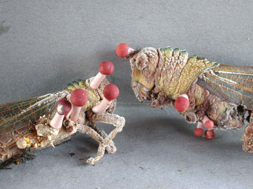
Somewhere in this thread, i have a post about parasitic fungi.
A specific kind of the cordyceps fungus is a parasitic fungi that takes over an ant.

In this youtube video it can be seen. As it turns out, there are a lot of cordyceps fungi (over 400 species), where each version of this fungus has it own favorite insect to prey upon.

http://www.youtube.com/watch?v=vgkL8PulPdE

http://en.wikipedia.org/wiki/Cordyceps

A fungi that has infected (i think it is) a locust.

Cordyceps_amazonica_9307TJB.jpg
 
May 11, 2008
19,469
1,159
126
I came across "the warburg effect".


I find this very interesting. What i understand of it, is that normally energy in the cell is produced by use of mitochondria. But in cancer cells , the cells can use another form of energy production. What kind of evolutionary leftover gene is responsible for this ability. It allows for cells to have more energy.

Can somebody explain the warburg effect in more detail to me ?
Are these qoutes correct ?

http://en.wikipedia.org/wiki/Warburg_effect

In oncology, the Warburg effect is the observation that most cancer cells predominantly produce energy by a high rate of glycolysis followed by lactic acid fermentation in the cytosol, rather than by a comparatively low rate of glycolysis followed by oxidation of pyruvate in mitochondria as in most normal cells.[4][5][6] The latter process is aerobic (uses oxygen). Malignant, rapidly growing tumor cells typically have glycolytic rates up to 200 times higher than those of their normal tissues of origin; this occurs even if oxygen is plentiful.

Otto Warburg postulated this change in metabolism is the fundamental cause of cancer,[7] a claim now known as the Warburg hypothesis. Today, mutations in oncogenes and tumor suppressor genes are known to be responsible for malignant transformation, and the Warburg effect is considered to be a result of these mutations rather than a cause.

http://medicalxpress.com/news/2014-08-reveals-cancer-energy-source.html

All multicellular organisms evolved pathways that take nutrients, sugars and oxygen and make energy through respiration and chemical processes. In normal cells, this energy-making process is known as oxidative phosphorylation. But when cells evolve cancerous properties and grow uncontrollably, they instead ferment their sugars to create energy even in the presence of oxygen. This process is called aerobic glycolysis, or the Warburg Effect.

The new findings are an important step toward developing a drug that affects only fermentation and not the normal metabolism of glucose, thereby depriving cancer cells of energy. The new model lays groundwork for predicting whether treatments will be effective based on an individual's unique metabolism.

Still, very few details have been known about the Warburg Effect. "We can now systematically perturb anything in the [computer] model and identify important components" of the Warburg Effect, Locasale said.

Dating back to work by Efraim Racker, a Cornell researcher who made seminal discoveries in the area in the 1970s, followed by advances in cancer and genetic research, it is "known now that almost every cancer gene has some capacity to induce the Warburg Effect," making it fundamental to proliferative diseases, Locasale said.

Currently, the Warburg Effect is used in clinical practice to diagnose and monitor cancer. Doctors inject patients with radioactive glucose and then watch where it is consumed; tumors are a major source of consumption. Researchers are also exploring whether dietary interventions with less sugar and the use of diabetes drugs that lower glucose may impact the Warburg Effect to treat cancer.
 
May 11, 2008
19,469
1,159
126
The big question is how life emerged from single celled organisms to multi cellular organisms. Multi cellular organisms where groups of cells take on a specific task. Nicole King might have found a clue. Once again it is clear that animal life would not have existed without bacteria.

I once opted for the fun that multi cellular life is beneficial to bacteria as we are a perfect transport medium to transport bacteria over vast distances, for example away from danger or to new sources of "food". Our bacterial masters. I am thinking about food rich but dangerous area such as near geysers, hydrothermal and vulcanoes.
Of course it is not that simple but one must admit that animal life is a handy transport vessels for bacteria and any other single and tiny organism like parasites and viruses.

Of course, bacteria can also start to work together and form large filaments or form sheets of cooperating bacteria. Bonnie Bassler has many interesting topics about cooperating bacteria and quorum sensing. To me it seems Salpingoeca rosetta also use a form of quorum sensing (by listening in) because when the right (Algoriphagus bacteria) bacteria is present and start using quorum sensing, S Rosetta also starts to form colonies.

http://en.wikipedia.org/wiki/Quorum_sensing


http://www.wired.com/2014/08/where-animals-come-from/


For billions of years, single-celled creatures had the planet to themselves, floating through the oceans in solitary bliss. Some microorganisms attempted multicellular arrangements, forming small sheets or filaments of cells. But these ventures hit dead ends. The single cell ruled the earth. Original story reprinted with permission from Quanta Magazine, an editorially independent division of SimonsFoundation.org whose mission is to enhance public understanding of science by covering research developments and trends in mathematics and the physical and life sciences.

Then, more than 3 billion years after the appearance of microbes, life got more complicated. Cells organized themselves into new three-dimensional structures. They began to divide up the labor of life, so that some tissues were in charge of moving around, while others managed eating and digesting. They developed new ways for cells to communicate and share resources. These complex multicellular creatures were the first animals, and they were a major success. Soon afterward, roughly 540 million years ago, animal life erupted, diversifying into a kaleidoscope of forms in what’s known as the Cambrian explosion. Prototypes for every animal body plan rapidly emerged, from sea snails to starfish, from insects to crustaceans. Every animal that has lived since then has been a variation on one of the themes that emerged during this time.

How did life make this spectacular leap from unicellular simplicity to multicellular complexity? Nicole King has been fascinated by this question since she began her career in biology. Fossils don’t offer a clear answer: Molecular data indicate that the “Urmetazoan,” the ancestor of all animals, first emerged somewhere between 600 and 800 million years ago, but the first unambiguous fossils of animal bodies don’t show up until 580 million years ago. So King turned to choanoflagellates, microscopic aquatic creatures whose body type and genes place them right next to the base of the animal family tree. “Choanoflagellates are to my mind clearly the organism to look at if you’re looking at animal origins,” King said. In these organisms, which can live either as single cells or as multicellular colonies, she has found much of the molecular toolkit necessary to launch animal life. And to her surprise, she found that bacteria may have played a crucial role in ushering in this new era.

Nicole King, a biologist at the University of California, Berkeley, studies the origins of animals, one of the big mysteries in the history of life. Courtesy of Nicole King

In a lengthy paper that will be published in a special volume of Cold Spring Harbor Perspectives in Biology in September, King lays out the case for the influence of bacteria on the development of animal life. For starters, bacteria fed our ancient ancestors, and this likely required those proto-animals to develop systems to recognize the best bacterial prey, and to capture and engulf them. All of these mechanisms were repurposed to suit the multicellular lives of the first animals. King’s review joins a broad wave of research that puts bacteria at the center of the story of animal life. “We were obliged to interact intimately with bacteria 600 million years ago,” said King, now an evolutionary biologist at the University of California, Berkeley, and an investigator with the Howard Hughes Medical Institute. “They were here first, they’re abundant, they’re dominant. In retrospect we should’ve expected this.”

Multicellular Motivation

Although we tend to take the rise of animals for granted, it is reasonable to ask why they ever emerged at all, given the billions of years of success of unicellular organisms. “For the last 3.5 billion years, bacteria have been around and abundant,” said Michael Hadfield, a professor of biology at the University of Hawaii, Manoa. “Animals never showed up until 700 or 800 million years ago.”

The technical demands of multicellularity are significant. Cells that commit to living together need a whole new set of tools. They have to come up with ways of sticking together, communicating, and sharing oxygen and food. They also need a master developmental program, a way to direct specific cells to take on specialized jobs in different parts of the body.

Nonetheless, during the course of evolution, the transition to multicellularity happened separately as many as 20 different times in lineages from algae to plants to fungi. But animals were the first to develop complex bodies, emerging as the most dramatic example of early multicellular success.

To understand why this might have happened the way it did, King began studying choanoflagellates, the closest living relative to animals, nearly 15 years ago as a postdoc at the University of Wisconsin, Madison. Choanoflagellates are not the most charismatic of creatures, consisting of an oval blob equipped with a single taillike flagellum that propels the organism through the water and also allows it to eat. The tail, thrashing back and forth, drives a current across a rigid, collarlike fringe of thin strands of cell membrane. Bacteria get caught up in the current and stick to the collar, and the choano engulfs them.

What intrigued King about choanoflagellates was their lifestyle flexibility. While many live as single cells, some can also form small multicellular colonies. In the species Salpingoeca rosetta, which lives in coastal estuaries, the cell prepares to divide but stops short of splitting apart, leaving two daughter cells connected by a thin filament. The process repeats, creating rosettes or spheres containing as many as 50 cells in the lab. If this all sounds familiar, there’s a reason for it — animal embryos develop from zygotes in much the same way, and spherical choanoflagellate colonies look uncannily like early-stage animal embryos.

When King began studying S. rosetta, she couldn’t get the cells to consistently form colonies in the lab. But in 2006, a student stumbled on a solution. In preparation for genome sequencing, he doused a culture with antibiotics, and it suddenly bloomed into copious rosettes. When bacteria that had been collected along with the original specimen were added back into a lab culture of single choanoflagellates, they too formed colonies. The likely explanation for this phenomenon is that the student’s antibiotic treatment inadvertently killed off one species of bacteria, allowing another that competes with it to rebound. The trigger for colony formation was a compound produced by a previously unknown species of Algoriphagus bacteria that S. rosetta eats.

S. rosetta seems to interpret the compound as an indication that conditions are favorable for group living. King hypothesizes that something similar could have happened more than 600 million years ago, when the last common ancestor of all animals started its fateful journey toward multicellularity. “My suspicion is that the progenitors of animals were able to become multicellular, but could switch back and forth based on environmental conditions,” King said. Later, multicellularity became fixed in the genes as a developmental program.

King’s persistence in studying this humble organism, which was overlooked by most contemporary biologists, has won her the admiration of many of her fellow scientists (as well as a prestigious MacArthur fellowship). “She strategically picked an organism to gain insight into early animal evolution and systematically studied it,” said Dianne Newman, a biologist at the California Institute of Technology in Pasadena, who studies how bacteria coevolve with their environment. King’s research offers a thrilling glimpse into the past, a rare window into what might have been going on during that mysterious period before the first fossilized animals appeared. The research is a “beautiful example” of how bacteria shape even the simplest forms of complex life, Newman said. “It reminds us that even at that level of animal development, you can expect triggers from the microbial world.” The bacteria system in S. rosetta can now be used to answer more specific questions, such as what the benefit of multicellularity might be — a question King and her collaborators at Berkeley are now working to answer.

The first bacteria may date back as far as 3.5 billion years. But animals, the first complex multicellular life form, took much longer to emerge. Russell Chun for Quanta Magazine

Of course, just because bacteria trigger modern choanoflagellates into group living, that doesn’t mean they had the same effect on the first proto-animals. King’s finding is “really cool,” said William Ratcliff, a biologist at the Georgia Institute of Technology in Atlanta who experimentally induces yeast to form multicellular colonies. “I think she’s doing some of the most interesting research in the origins of animals.” But, he cautions, it’s possible that choanoflagellates evolved this mechanism long after they diverged from the creatures that became the first ancestors of animals. “We don’t have a clear picture of when the bacterial response evolved,” he explained. “It’s hard to know if something happened before the split between choanoflagellates and animals, or after.”

“I think there is enough evidence to allow us to hypothesize that bacteria were an important influence on animal origins — they were abundant, diverse, and they exert important signaling influences on diverse animal lineages as well as on non-animals,” King said. “But I think it is premature to say what the nature of that influence was.”

One strong hint that bacteria may have prompted that ancient transition to multicellularity is that many of today’s simplest animals are governed by microbial messages. Corals, sea squirts, sponges and tube worms all begin life as larvae floating in the water, and other research teams have shown that they too respond to compounds released by bacteria as signals to attach themselves to rocks or other surfaces and transition to a new life form. If this kind of relationship is so common among animals from the most ancient families, it seems plausible that the first animals were equally attuned to their bacterial neighbors. Figuring out how, exactly, the bacteria trigger this response will help clarify whether they played a similar role long ago. “It was a radical thought to me when we first started studying it, and now I don’t know why it’s a surprise,” King said. “The more I think about host-microbe interactions, the less surprised I become.”

What Took Animals So Long?

What triggered the explosion of complex multicellular life in the Cambrian period? Increased oxygen undoubtedly had something to do with it — prior to a period sometime before 800 million years ago, atmospheric oxygen levels were too low to diffuse easily into organisms with multiple layers of cells, limiting the size of all life forms. But an increase in oxygen is probably not the whole story, said Andrew Knoll, a professor of earth and planetary sciences at Harvard University. Once oxygen levels rose past this low level, predation likely provided a strong incentive for animals to get bigger and more complicated, and to develop new body plans. It was an ecological arms race of size and complexity: Bigger predators have an advantage in catching prey, while larger prey can more easily avoid being eaten. The need to escape or repel predators also likely inspired the first scales, spines and body armor, as well as some of the wilder body plans seen in Cambrian fossils.


King’s discovery about choanoflagellates is just one of the latest insights into the intimate relationships between bacteria and animals (or, in this case, animal-like organisms). Historically, photosynthetic bacteria pumped oxygen into the oceans for billions of years, setting the stage for complex multicellular life. And according to the endosymbiotic theory, proposed in the 20th century and now widely accepted, the mitochondria inside every eukaryotic cell were once free-living bacteria. At some point more than a billion years ago, they took up residence inside other cells in a symbiotic relationship that endures in nearly every animal cell to this day. In their role as dinner, bacteria also likely provided raw genetic material for the first animals, which probably incorporated chunks of microbial DNA directly into their own genomes as they digested their meals.

But the full story of the microbial-animal relationship is even broader and deeper, argues Margaret McFall-Ngai, a biologist at the University of Wisconsin, Madison, and it’s a story that is only beginning to be told. In her view, animals should rightly be considered host-microbe ecosystems. Several years ago McFall-Ngai, along with Hadfield, convened a broad group of developmental biologists, ecologists, environmental biologists and physiologists, including King, and asked them to formulate a microbial manifesto — a declaration of bacterial significance. The paper, which appeared late last year in the Proceedings of the National Academy of Sciences, cites evidence from many corners of biology to argue that the influence of microbes on the origin, evolution and function of animals is pervasive and essential to understanding how animal life evolved. “They evolved in a world saturated with bacteria,” Hadfield said.

The biology of choanoflagellates resembles that of animals in other unexpected ways, King found. In 2008 she led the team that published the genome of Monosiga brevicollis, a choanoflagellate that doesn’t form colonies. The sequence revealed genes for dozens of sections of proteins that also appear in multicellular animals, where they help cells stick together and also guide development and differentiation. What are they doing in single cells? King’s work suggests they arose in single-celled organisms to monitor environmental conditions and recognize other cells such as bacterial prey. In multicellular animals, the gene domains found new purposes, such as allowing cells to signal one another. Single cells used these tools to listen in on the environment. Later on, the first cells to adopt a multicellular lifestyle probably repurposed the same systems to pay attention to their sister cells, King suggested.

The breadth and significance of the animal-bacteria relationship goes far beyond the development of a handful of ancient aquatic creatures like sponges. McFall-Ngai’s own research shows that bacteria are necessary for the development of organs in squid; others have found similar partnerships that shape the maturation of animal immune systems, the guts of zebra fish and mice, and even mammalian brains. Likewise, bacteria are essential partners in the digestive systems of creatures ranging from termites to humans. The influence of microbes is even inscribed on our genome: More than a third of human genes have their origins in bacteria. These and other new findings will soon fundamentally alter our understanding of life, McFall-Ngai predicts: “Biology is in a revolution.”

So in the end, maybe animals really aren’t all that special. After all, they’d be nothing without their microbial friends. And as King’s research has revealed, much of what animals do that seems to make them interesting can also be accomplished by choanoflagellates. To her, that doesn’t diminish either one. “I love choanoflagellates,” she said. “They’re so fascinating. I see that they’re doing a lot of the same things as animals, and I can see parallels between their biology and the cell biology of animals. I could watch them for hours.”
 
Last edited:
May 11, 2008
19,469
1,159
126
Recently there was an outbreak of a bacteria genus called Listeria in Denmark.
This bacteria can causes serious illness and even death because of sepsis and meningitis.
Sepsis is put simple, inflammation of the entire body.
Meningitis is put simple, inflammation of the brain and or the central nervous system, the spinal cord.

http://en.wikipedia.org/wiki/Listeria


Very interesting stuff :

Listeria uses the cellular machinery to move around inside the host cell: It induces directed polymerization of actin by the ActA transmembrane protein, thus pushing the bacterial cell around.[10]

Listeria monocytogenes, for example, encodes virulence genes that are thermoregulated. The expression of virulence factor is optimal at 39 °C, and is controlled by a transcriptional activator, PrfA, whose expression is thermoregulated by the PrfA thermoregulator UTR element. At low temperatures, the PrfA transcript is not translated due to structural elements near the ribosome binding site. As the bacteria infect the host, the temperature of the host melts the structure and allows translation initiation for the virulent genes.

The majority of Listeria bacteria are targeted by the immune system before they are able to cause infection. Those that escape the immune system's initial response, however, spread through intracellular mechanisms and are, therefore, guarded against circulating immune factors (AMI).[8]

To invade, Listeria induces macrophage phagocytic uptake by displaying D-galactose in their teichoic acids that are then bound by the macrophage's polysaccharide receptors. Other important adhesins are the internalins.[9] Once phagocytosed, the bacterium is encapsulated by the host cell's acidic phagolysosome organelle.[7] Listeria, however, escapes the phagolysosome by lysing the vacuole's entire membrane with secreted hemolysin,[11] now characterized as the exotoxin listeriolysin O.[7] The bacteria then replicate inside the host cell's cytoplasm.[8]

Listeria must then navigate to the cell's periphery to spread the infection to other cells. Outside the body, Listeria has flagellar-driven motility, sometimes described as a "tumbling motility". However, at 37 °C, flagella cease to develop and the bacterium instead usurps the host cell's cytoskeleton to move.[8] Listeria, inventively, polymerizes an actin tail or "comet",[11] from actin monomers in the host's cytoplasm [6] with the promotion of virulence factor ActA.[8] The comet forms in a polar manner [12] and aids the bacteria's migration to the host cell's outer membrane. Gelsolin, an actin filament severing protein, localizes at the tail of Listeria and accelerates the bacterium's motility.[12] Once at the cell surface, the actin-propelled Listeria pushes against the cell's membrane to form protrusions called filopods[7] or "rockets". The protrusions are guided by the cell's leading edge [13] to contact adjacent cells, which then engulf the listeria rocket and the process is repeated, perpetuating the infection.[8] Once phagocytosed, the bacterium is never again extracellular: it is an intracytoplasmic parasite [11] like Shigella flexneri and Rickettsia.[8]

Thermoregulation, that is interesting stuff.
Interesting that that works best for this bacteria at a temperature that is common when the host (human) is having a fever.
 
Last edited:

Gibsons

Lifer
Aug 14, 2001
12,530
35
91
I came across "the warburg effect".


I find this very interesting. What i understand of it, is that normally energy in the cell is produced by use of mitochondria. But in cancer cells , the cells can use another form of energy production. What kind of evolutionary leftover gene is responsible for this ability. It allows for cells to have more energy.

Can somebody explain the warburg effect in more detail to me ?
Are these qoutes correct ?

Normal cells will use mitochondria (oxidative phosphorylation process) and/or glycolysis for energy. Some cells will use one or the other more often, it really depends on a lot of things. Ox-phos is generally a more efficient way to get ATP from glucose and requires oxygen, while glycolysis is faster (sort of) and doesn't require oxygen. Sprinters will use glycolysis, distance runners ox-phos.

Cancer cells use glycolysis predominantly or exclusively. To my knowledge, no one knows why. I'm sure there are some pseudoscience types who claim to know, but...

There's usually some speculation about the ability to survive in low oxygen environments, but the Warburg effect is maintained in cancers/cancer cells that have plenty of oxygen access. So I don't really buy into that, though it's probably important in quite a few cases. I tend to think it has something to do with the ability to avoid apoptosis - mitochondria are important players in apoptosis (see cytochrome C).
 
May 11, 2008
19,469
1,159
126
Normal cells will use mitochondria (oxidative phosphorylation process) and/or glycolysis for energy. Some cells will use one or the other more often, it really depends on a lot of things. Ox-phos is generally a more efficient way to get ATP from glucose and requires oxygen, while glycolysis is faster (sort of) and doesn't require oxygen. Sprinters will use glycolysis, distance runners ox-phos.

Cancer cells use glycolysis predominantly or exclusively. To my knowledge, no one knows why. I'm sure there are some pseudoscience types who claim to know, but...

There's usually some speculation about the ability to survive in low oxygen environments, but the Warburg effect is maintained in cancers/cancer cells that have plenty of oxygen access. So I don't really buy into that, though it's probably important in quite a few cases. I tend to think it has something to do with the ability to avoid apoptosis - mitochondria are important players in apoptosis (see cytochrome C).

Thank you. I will dig into it deeper. What you have written seems that it is more common that i thought. It seemed as i read something revolutionary when i was reading about the Warburg effect.
 
May 11, 2008
19,469
1,159
126
Normal cells will use mitochondria (oxidative phosphorylation process) and/or glycolysis for energy. Some cells will use one or the other more often, it really depends on a lot of things. Ox-phos is generally a more efficient way to get ATP from glucose and requires oxygen, while glycolysis is faster (sort of) and doesn't require oxygen. Sprinters will use glycolysis, distance runners ox-phos.

Cancer cells use glycolysis predominantly or exclusively. To my knowledge, no one knows why. I'm sure there are some pseudoscience types who claim to know, but...

There's usually some speculation about the ability to survive in low oxygen environments, but the Warburg effect is maintained in cancers/cancer cells that have plenty of oxygen access. So I don't really buy into that, though it's probably important in quite a few cases. I tend to think it has something to do with the ability to avoid apoptosis - mitochondria are important players in apoptosis (see cytochrome C).

I think you have a point. But is it also not the case that cancerous cells require lot of energy for the fast division ? And that glycolysis can provide that energy ?
Are the rates known about how fast different tumors can grow once out of control ? I read that people who do not eat much, also have less food to burn and that cancers develop more slowly in people with a moderate diet. However, people who consume a lot of calories when also developing a tumor, would have faster developing and growing tumors ? Do you have any knowledge of this , Gibsons ?

Cancer is really amazing in a scary way. It is almost as if the cell is trying to avoid death and wants to live forever. I wonder what the shortening of telomeres would mean for cancerous cells.
I always read in the past, that telomeres get shorter and that the cell cannot divide anymore at a certain moment. Now i read that the shortening of telomeres is reversed by by an enzyme, telomerase reverse transcriptase.

And here i found interesting stuff.
http://en.wikipedia.org/wiki/Telomere#Cancer
 

Gibsons

Lifer
Aug 14, 2001
12,530
35
91
I think you have a point. But is it also not the case that cancerous cells require lot of energy for the fast division ? And that glycolysis can provide that energy ?

Certainly, and yes.

Not all cancers grow quickly though. Generally, the faster they grow the more deadly they are, but that's also partly a product of how evaluation is done (e.g. 5 year survival data).

Are the rates known about how fast different tumors can grow once out of control ?

There are numbers, I'm not sure how accurate they are for real in vivo cases. And again, they don't all grow fast.

I read that people who do not eat much, also have less food to burn and that cancers develop more slowly in people with a moderate diet. However, people who consume a lot of calories when also developing a tumor, would have faster developing and growing tumors ? Do you have any knowledge of this , Gibsons ?

Don't know, but it wouldn't surprise me.

Cancer is really amazing in a scary way. It is almost as if the cell is trying to avoid death and wants to live forever. I wonder what the shortening of telomeres would mean for cancerous cells.

Forget 'trying.' They're just cells. And, like any any imperfect replicator, they evolve.

Still being worked on of course, but the ones that are good at being cancer are the ones we see. Some phenotypes: lack of apoptosis, increased growth rate, immune avoidance, ability to move and colonize new territory etc..... and probably some things we don't know about or haven't considered.
I always read in the past, that telomeres get shorter and that the cell cannot divide anymore at a certain moment. Now i read that the shortening of telomeres is reversed by by an enzyme, telomerase reverse transcriptase.
And here i found interesting stuff.
http://en.wikipedia.org/wiki/Telomere#Cancer

Yes, active telomerase is a common feature, it's formally required for immortilization afwk.
I was at a talk by Elizabeth Blackburn about a year ago, and she had some evidence that there was more to telomerase than just reverse transcription. I'm fuzzy on the details and too lazy to look it up atm.
 
Last edited:
May 11, 2008
19,469
1,159
126
Certainly, and yes.

Forget 'trying.' They're just cells. And, like any any imperfect replicator, they evolve.

Of course you are right about "trying" , bad choice of words.
I just mean to write that these cells evolved from a typical cell in the body to a cell that can seemingly live forever. But of course, there comes a point that some of those cancer cells evolve into a cell that just stops functioning. But some might evolve into a cell that can continue to live as long as there is an environment to sustain them.

I read that when researchers looked at a tumor, the tumor had lot's of differentiated cells. Each starting to evolve into a different function. Not really evolved into a new kind of organism, it was more that some of these these cells showed different activities compared to others in the tumor.

And there is one form of cancer that i find very interesting and is described here in this thread. It is a parasitic cancer :

http://forums.anandtech.com/showpost.php?p=30237562&postcount=130
Just a strange question. But is Herpes simplex virus not infecting Schwann cells ?

I was wondering about the coincidence between the Devil facial tumour disease
in tasmanian devils. This disease is an parasitic cancer. And the hypothesis is that it originated inside schwann cells.

Parasitic cancers or transmissible cancers are very rare but i find it very interesting that the immunesystem from tasmanian devils does not seem to mind that an foreign cell can just start to grow like a tumor. Perhaps this has something to do with the fact that tasmanian devils are isolated as an species. Interbreeding might occur and may cause unwanted side effects as for example that the immune system becomes dumbed down. I do not know.


http://en.wikipedia.org/wiki/Devil_facial_tumour_disease

http://en.wikipedia.org/wiki/Parasitic_cancer

This cancer likely evolved from being just a cancer to a transmittable form of cancer. But i still wonder if the start was some kind of pathogen infection.
 
Last edited:
May 11, 2008
19,469
1,159
126
What is really scary but luckily very rare and only happens with the syrian hamster is a form of parasitic cancer that is transmitted from hamster to hamster by a mosquito bite.

This is from research done in 1967.
http://www.ncbi.nlm.nih.gov/pmc/articles/PMC1841392/
Transfer of Tumour Cells by Mosquitoes

Until a few years ago the suggestion that'mosquitoes might transmit cancer would have been treated with scepticism. But now there is a suspicion that certain species may transmit the virus or viruses thought to cause Burkitt's lymphoma.' 2
Moreover, W. G. Banfield and his colleagues2 ' have shown that mosquitoes can also transmit a lymphoma in experimental animals. The mechanisms are of considerable interest,particularly as they appear to be radically different from those believed to operate in the Burkitt tumour. These experimental studies were carried out on a reticulum cell sarcoma which originally occurred as a single, apparently spontaneous, neoplasm in an old hamster.' This tumour is readily transplanted by conventional techniques, grows rapidly at the site of transplantation, and metastasizes widely, with a late leukaemic phase. Mosquitoes (Aides aegypti) allowed to feed on animals during the leukaemic stage proved capable of transferring the lymphoma to normal hamsters.2'
Two techniques were employed to demonstrate this. In the first, mosquitoes in the post-prandial state were placed subcutaneously in normal animals and then crushed; in the second, the mosquitoes were allowed to bite the ecipient animals. Although the incidence of positive "takes" was much lower by the latter method (10% as opposed to 88%), both groups showed one remarkable feature-the tumour was still transmissible for up to eight hours after the mosquito had fed. In all instances in which the tumour was successfully transferred the clinical course of the disease was indistinguishable from that observed in the original donor animals. These are intriguing observations, but many features are still obscure. The role of the mosquito is probably purely passive and the actual species may be irrelevant. Nevertheless, it seems desirable to check this point by comparing the performance of a number of different species. Next, there is the apparent resistance of reticulum cell sarcoma cells to mechanical and chemical damage during their sojourn in the mosquito. Their distribution is uncertain, but they are clearly not confined to the mouth parts and proximal portions of the foregut. Some lymphoma cells may accumulate in the dorsal and ventral diverticula, but Banfield and his colleagues described large numbers of tumour cells in the midgut,3 the main site of secretion of digestive enzymes.5 Unless the tumour cells are unusually resistant to these enzymes many must surely be destroyed. This implies that successful "takes " can be achieved by the transference of relatively few viable tumour cells, but more information on this point, using standard quantitative transplantation methods, is clearly needed. On the other hand, there are grounds for supposing that the tumour cells are indeed unusually robust, in so far as the tumour has apparently been successfully transmitted between hamsters by simple feeding.4 Confirmation of this remarkable finding would seem to be desirable, preferably combined with some in-vitro studies on the cells, in an attempt to clarify the nature of their extraordinary resistance. The exact sequence of events which takes place when mosquitoes bite the hamsters is obscure. It is not clear, for example, whether tumour cells are transferred in the saliva or whether viable cells are also regurgitated from lower down the gut, particularly from the foregut diverticula. Finally, there is the problem of what exactly is transmitted-tumour
cells only, a virus only, or tumour cells plus virus ? No tumours have been induced with a wide range of cell-free filtrates,4 and the authors emphasize the unusually consistent karyotype shown by the tumour6; seven extra chromosomes are regularly present, including a distinctive marker chromosome. This is in contrast to the more unstable karyotype
patterns seen in cells transformed by viruses such as SV 407 and polyoma.8 These facts support the view that transference was by cells in the present experiments, but they do not exclude the possibility that the reticulum cell tumour was originally induced by a virus. Though much remains to be clarified, it appears that in certain circumstances the mosquito may transmit enough viable tumour cells to healthy recipients to induce tumours in them. But undoubtedly the lymphoma used by Banfield and his colleagues is highly unusual and the relevance of their findings to the transmission of the Burkitt tumour and other human cancers cannot yet be assessed. Further information will be awaited with interest.

* Stanley, N. F., Lancet, 1966, 1, 961.
Banfield, W. G., Woke, P. A., MacKay, C. M., and Cooper, H. L.,
Science, 1965, 148, 1239.
'-- Cancer (Philad.), 1966, 19, 1333.
Brindley, D. C., and Banfield, W. G., 7. nat. Cancer Inst., 1961, 26,
949.
' Clements, A. N., The Physiology of Mosquitoes, 1963. Oxford.
' Cooper, H. L., MacKay, C. M., and Banfield, W. G., 7. nat. Cancer
Inst., 1964, 33, 691.
Macpherson, I., ibid., 1963, 30, 795.
Cooper, H. L., and Black, P. H., ibid., 30, 1015.
 
May 11, 2008
19,469
1,159
126
Today i was at the pharmacist, and while i was waiting for my recipe, i was reading about cancer immunotherapy. This therapy is experimental and expensive but has good results. Simply put, the cancer cells produce proteins that suppress T cells. and in this therapy, T cells are modified, cultivated and placed back inside the body of the cancer patient to seek out and destroy cancer cells, these T-cells attack the cancer cells . Now this has some serious side effects, Some of these T-Cells will attack anything and a lot of cancer patients receiving the therapy (Who obviously would have died otherwise) start to exhibit auto-immune diseases as side effect. But this can be suppressed by modern medicines. This is a great advancement. The whole idea behind it is to let the body fight the cancer off. But cancer cells evolve strategies to prevent from being destroyed. The human research conquest is now to learn T cells to not be fooled by cancer cells. And this research is advancing.

http://www.sciencemag.org/content/342/6165/1432.full
Short version :
Science's editors have chosen cancer immunotherapy as Breakthrough of the Year for 2013, a strategy that harnesses the body's immune system to combat tumors. It's an attractive idea, and researchers have struggled for decades to make it work. Now, many oncologists say those efforts are paying off, as two different techniques show signs of helping some patients. One involves antibodies that release a brake on T cells, giving them the power to tackle tumors. Another involves genetically modifying an individual's T cells outside the body to make them better able to target cancer, and then reinfusing them so they can do just that. Experts stress that these techniques have been tested in only small trials, and they don't always work. But the results have raised hope that immunotherapy may give doctors new options for treatment in the future.

Long version :
History's path is unchartable when it's not yet past but present, when we are still standing in the middle of it. That's what made Science's selection of this year's Breakthrough of the Year such a topic of internal debate, even anxiety. In celebrating cancer immunotherapy—harnessing the immune system to battle tumors—did we risk hyping an approach whose ultimate impact remains unknown? Were we irresponsible to label as a breakthrough a strategy that has touched a tiny fraction of cancer patients and helped only some of them? What do we mean when we call something a breakthrough, anyway?

Ultimately, we concluded, cancer immunotherapy passes the test. It does so because this year, clinical trials have cemented its potential in patients and swayed even the skeptics. The field hums with stories of lives extended: the woman with a grapefruit-size tumor in her lung from melanoma, alive and healthy 13 years later; the 6-year-old near death from leukemia, now in third grade and in remission; the man with metastatic kidney cancer whose disease continued fading away even after treatment stopped.

As the anecdotes coalesce into data, there's another layer, too, a sense of paradigms shifting. Immunotherapy marks an entirely different way of treating cancer—by targeting the immune system, not the tumor itself. Oncologists, a grounded-in-reality bunch, say a corner has been turned and we won't be going back.

With much pressure these days to transform biological insights into lifesaving drugs, there's a lesson to be learned from immunotherapy's successes: They emerged from a careful decoding of basic biology that spanned many years. The early steps were taken by cancer immunologist James Allison, now at the University of Texas MD Anderson Cancer Center in Houston. In the late 1980s, French researchers who weren't thinking about cancer at all identified a new protein receptor on the surface of T cells, called cytotoxic T-lymphocyte antigen 4, or CTLA-4. Allison found that CTLA-4 puts the brakes on T cells, preventing them from launching full-out immune attacks. He wondered whether blocking the blocker—the CTLA-4 molecule—would set the immune system free to destroy cancer.

Allison's rationale was untested. He and his colleagues changed the conversation, in the words of one cancer researcher, "to consider immunosuppression as the focal point, and manipulation of immunosuppression as the target."

Doing so took time. CTLA-4 was discovered in 1987. In 1996, Allison published a paper in Science showing that antibodies against CTLA-4 erased tumors in mice. Pharmaceutical companies shied away from cancer immunotherapy, wary of past flops but also of a strategy very unlike the standard zapping of a tumor. So the job of getting anti–CTLA-4 into people fell to a small biotechnology company, Medarex, in Princeton, New Jersey. In 1999, it acquired rights to the antibody, taking the leap from biology to drug.

Crucial results didn't come for another 11 years. In 2010, Bristol-Myers Squibb—which had bought Medarex for more than $2 billion—reported that patients with metastatic melanoma lived an average of 10 months on the antibody, compared with 6 months without it. It was the first time any treatment had extended life in advanced melanoma in a randomized trial. Nearly a quarter of participants survived at least 2 years.

The numbers for another antibody are so far even better and the side effects milder. In the early 1990s, a biologist in Japan discovered a molecule expressed in dying T cells, which he called programmed death 1, or PD-1, and which he recognized as another brake on T cells. He wasn't thinking of cancer, but others did. One, oncologist Drew Pardoll at Johns Hopkins University, met with a leader of Medarex at a Baltimore coffee shop. He urged the company to test an anti–PD-1 antibody in people.

The first trial, with 39 patients and five different cancers, began in 2006. By 2008, doctors were jolted by what they saw: In five of the volunteers, all of them with refractory disease, tumors were shrinking. Survival in a few stretched beyond what was imagined possible.

Still, understanding what these therapies were doing inside the body was a challenge. Other cancer treatments either work or they don't, and the answer is nearly instantaneous. With both anti–CTLA-4 and anti–PD-1, physicians saw some tumors grow before vanishing months later. Some patients kept responding even after the antibody had been discontinued, suggesting their immune system had been fundamentally changed. Some, particularly those on anti–CTLA-4, developed unnerving side effects, inflammation of the colon, for example, or of the pituitary gland. All of these were the fine points of a new template, one whose vagaries physicians were just beginning to understand. The learning curve would be steep.

It was steep in another area of immunotherapy as well. For years, Steven Rosenberg at the National Cancer Institute had harvested T cells that had migrated into tumors, expanded them in the lab, and reinfused them into Tpatients, saving some with dire prognoses. The technique worked only when doctors could access tumor tissue, though, limiting its application.

Then in 2010, Rosenberg published encouraging results from so-called chimeric antigen receptor therapy, or CAR therapy—a personalized treatment that involves genetically modifying a patient's T cells to make them target tumor cells. One group, led by Carl June at the University of Pennsylvania, began reporting eye-catching responses to CAR therapy: patients with pounds of leukemia that melted away. At a meeting in New Orleans this month, June's team and another at Memorial Sloan-Kettering Cancer Center in New York reported that the T cell therapy in their studies put 45 of 75 adults and children with leukemia into complete remission, although some later relapsed. CAR therapy is now the focus of numerous clinical trials. Researchers hope that it, like the antibodies, can target an assortment of cancers.


This Year's Breakthrough and Runners Up

Engineered T cells are still experimental, but the antibodies are slowly going mainstream. At least five major drug companies, their early hesitancy gone, are developing antibodies such as anti–PD-1. In 2011, the U.S. Food and Drug Administration approved Bristol-Myers Squibb's anti–CTLA-4 treatment, called ipilimumab, for metastatic melanoma. The cost is high: The company charges $120,000 for a course of therapy. In 2012, Suzanne Topalian of Hopkins, Mario Sznol of Yale University, and their colleagues reported results for anti–PD-1 therapy in nearly 300 people, and they provided an update earlier this year. Tumors shrunk by about half or more in 31% of those with melanoma, 29% with kidney cancer, and 17% with lung cancer.

This year brought even more encouragement. Bristol-Myers Squibb reported this fall that of 1800 melanoma patients treated with ipilimumab, 22% were alive 3 years later. In June, researchers reported that combining ipilimumab and anti–PD-1 led to "deep and rapid tumor regression" in almost a third of melanoma patients. Drugs blocking the PD-1 pathway have not yet been proven to extend life, although survival rates so far have doctors optimistic that they do.

For physicians accustomed to losing every patient with advanced disease, the numbers bring a hope they couldn't have fathomed a few years ago. For those with metastatic cancer, the odds remain long. Today's immunotherapies don't help everyone, and researchers are largely clueless as to why more don't benefit. They are racing to identify biomarkers that might offer answers and experimenting with ways to make therapies more potent. It's likely that some cancers will not yield to immunotherapy for many years, if ever.

Even in the fluid state oncology now finds itself, this much is certain: One book has closed, and a new one has opened. How it will end is anyone's guess.

EDIT :
I was thinking that using this therapy first and then a milder form of chemotherapy would also have good results. Or first a milder form of chemotherapy and then immunotherapy as the finishing touch.
 
Last edited:
May 11, 2008
19,469
1,159
126
Anyone who reads this thread, might get the impression that we are saturated with bacteria and other micro organisms. Especially when reading the posts about Bonnie Bassler.
The next idea however might be that we spread our bacteria in the environment we are in. And as it turns out, this is the case. I am willing to bet that each human has a sort of unique microorganism fingerprint. And that that bacterial aura also affects the host (for example a human). I should also note that the micro organism environment a person resides in will also influence that person over time. Meaning the friendly "body" bacteria and the immune system (symbiotic) are once again fighting off invaders for a place to live on this planet.

Researchers have found that this bacterial aura follows a person around wherever that person resides for a while. And i was willing to bet in the past it is family specific ( which turns out to be the case). So i would also not be surprised to find out that some family "diseases" or genetic diseases are also partial explainable because of the micro organisms they carry along with them.
Diseases that are explainable through the effects of the gut on the body. But in the nasal cavities and the sinuses, there is also a bacterial culture residing.

http://blogs.discovermagazine.com/d-brief/2014/08/28/microbiome-bacterial-aura-follows-you/

Researchers from the U.S. Department of Energy’s Argonne National Laboratory and the University of Chicago have found that the communities of microbes living in individual homes are unique and identifiable, meaning humans sharing a home have a similar microbial “aura.” And, these microbial passengers follow us wherever we go, swiftly populating new homes, and even hotel rooms, within less than a day of occupancy.

Under the Influence
The study was conducted as a part of the Home Microbiome Project, which aims to understand how humans interact with, and influence, the bacteria in their homes.
The bugs were collected from seven U.S. families (comprising 18 people, three dogs and one cat), who dutifully swabbed their homes and themselves every day over the course of six weeks. Samples included microbes from hands, feet and noses, as well as doorknobs, light switches, floors and counter-tops.
The study subjects were ethnically diverse, and not all occupants in individual homes were genetically related. Three of the families moved to new homes during the course of the study – one of them from a hotel room.
By performing DNA analysis on these daily swabs, researchers were able to characterize the different species of microbes within each dwelling, and how they changed over time.
Sharing is Caring
Millions of bacteria were sequenced and identified from the project. The researchers found that every home has a unique bacterial fingerprint, which follows families when they move. When comparing the surfaces of the old and new homes, researchers found them almost identical in microbial makeup – suggesting that our bacteria settle down in new places before our boxes are even unpacked. What’s more, a household’s fingerprint is unique enough that the researchers could use an unidentified floor sample to predict which of the seven families it came from.
Within a household, hands were most likely to contain similar bacteria, while noses showed the most variation. Pets made a significant contribution to the house’s microbial makeup, in line with previous research that found that two strangers who own dogs share as many microbes as people cohabiting do.
Researchers also found that physical contact may influence your microbial makeup. In one home, none of the occupants were genetically related. But, the two occupants who were in a relationship shared more bacteria than the third housemate. And, parents and their children have markedly similar microbial makeups. The results are published in Science this week.
Making Your Mark
Through our microbes, then, it seems we could be leaving more of a mark on our surroundings than we ever imagined – which the researchers say could prove useful for forensics in the future.
In their study, when someone departed his or her house for a few days, researchers could discern that person’s absence in the microbial aura. And by piecing together samples from different surfaces, researchers say they could pin down a pretty precise chronology of comings and goings within a house.
So, next time you sit down to relish the calm of a clean house, remember that you’re not quite alone – nor would you want to be.
 
Last edited:
May 11, 2008
19,469
1,159
126
This thread started with bacteriophages and is not going to end with a post about phages. Since bacteria become increasingly resistant against antibiotics and the research into phages is going on steady, there is now good news for disinfecting medical implants.


http://phys.org/news/2013-05-bacteria-eaters-infections-medical-implant-materials.html#nRlv

usingbacteri.gif


They're ba-ack! But in a new disease-fighting role. Viruses that infect and kill bacteria—used to treat infections in the pre-antibiotic era a century ago and in the former Soviet Union today—may have a new role in preventing formation of the sticky "biofilms" of bacteria responsible for infections on implanted medical devices. That's the topic of a report in the ACS journal Biomacromolecules.

Marek Urban and colleagues explain that bacteriophages (literally, "bacteria eaters") were first used to treat bacterial infections in the 19th century. These viruses—more than 1,000 different kinds exist—attack disease-causing bacteria. The scientists focused on use of phages to wage "microbial warfare" on the films of bacteria that form on catheters, stents and other medical implants. These infections, which often involve antibiotic-resistant bacteria, strike more than a million patients annually in the United States alone, increasing hospital bills by almost $1 billion.

They describe attachment of phages to the surfaces of materials like those used in implanted medical devices, and evidence that the phages remain active, killing E. coli and Staphylococcus aureus. Those bacteria cause the most common hospital-acquired infections. The technology can attach phages to almost any surface, and is "a promising and effective means of not only combating antibiotic-resistant infections, but also the technological platform for the development of bacteria sensing and detecting devices."
 
May 11, 2008
19,469
1,159
126
There has been research done where it was found out that neurons in the brain either have specific receptors for artificial sweeteners or acquire specific receptors. And there is the ongoing debate if artificial sweeteners are more damaging than their natural counterparts.

Recent research seems to suggests that once again our gut bacteria may play a role in many aspects of our life. One such example is research that seems to suggest that gut bacteria play a key role in a controlling and boosting the immune system after a vaccine against the flu is medicated. As it turns out, the bacteria in the gut play a key role in how strong the immune system of a person is. With all the posts about bacteria in this thread, this evidence is stacking. Of course, it depends on specific species of bacteria. It is not the case that any random bacteria will be beneficial. As always, it is in the numbers and the composition of different species of bacteria living in the gut.

But there is more...

http://www.sciencedaily.com/releases/2014/09/140917131634.htm

Certain gut bacteria may induce metabolic changes following exposure to artificial sweeteners

Artificial sweeteners -- promoted as aids to weight loss and diabetes prevention -- could actually hasten the development of glucose intolerance and metabolic disease, and they do so in a surprising way: by changing the composition and function of the gut microbiota -- the substantial population of bacteria residing in our intestines. These findings, the results of experiments in mice and humans, were published September 17 in Nature. Dr. Eran Elinav of the Weizmann Institute of Science's Department of Immunology, who led this research together with Prof. Eran Segal of the Department of Computer Science and Applied Mathematics, says that the widespread use of artificial sweeteners in drinks and food, among other things, may be contributing to the obesity and diabetes epidemic that is sweeping much of the world.


For years, researchers have been puzzling over the fact that non-caloric artificial sweeteners do not seem to assist in weight loss, with some studies suggesting that they may even have an opposite effect. Graduate student Jotham Suez in Dr. Elinav's lab, who led the study, collaborated with lab member Gili Zilberman-Shapira and graduate students Tal Korem and David Zeevi in Prof. Segal's lab to discover that artificial sweeteners, even though they do not contain sugar, nonetheless have a direct effect on the body's ability to utilize glucose. Glucose intolerance -- generally thought to occur when the body cannot cope with large amounts of sugar in the diet -- is the first step on the path to metabolic syndrome and adult-onset diabetes.

The scientists gave mice water laced with the three most commonly used artificial sweeteners, in amounts equivalent to those permitted by the U.S. Food and Drug Administration (FDA). These mice developed glucose intolerance, as compared to mice that drank water, or even sugar water. Repeating the experiment with different types of mice and different doses of the artificial sweeteners produced the same results -- these substances were somehow inducing glucose intolerance.

Next, the researchers investigated a hypothesis that the gut microbiota are involved in this phenomenon. They thought the bacteria might do this by reacting to new substances like artificial sweeteners, which the body itself may not recognize as "food." Indeed, artificial sweeteners are not absorbed in the gastrointestinal tract, but in passing through they encounter trillions of the bacteria in the gut microbiota.

The researchers treated mice with antibiotics to eradicate many of their gut bacteria; this resulted in a full reversal of the artificial sweeteners' effects on glucose metabolism. Next, they transferred the microbiota from mice that consumed artificial sweeteners to "germ-free," or sterile, mice -- resulting in a complete transmission of the glucose intolerance into the recipient mice. This, in itself, was conclusive proof that changes to the gut bacteria are directly responsible for the harmful effects to their host's metabolism. The group even found that incubating the microbiota outside the body, together with artificial sweeteners, was sufficient to induce glucose intolerance in the sterile mice. A detailed characterization of the microbiota in these mice revealed profound changes to their bacterial populations, including new microbial functions that are known to infer a propensity to obesity, diabetes, and complications of these problems in both mice and humans.

Does the human microbiome function in the same way? Dr. Elinav and Prof. Segal had a means to test this as well. As a first step, they looked at data collected from their Personalized Nutrition Project (www.personalnutrition.org), the largest human trial to date to look at the connection between nutrition and microbiota. Here, they uncovered a significant association between self-reported consumption of artificial sweeteners, personal configurations of gut bacteria, and the propensity for glucose intolerance. They next conducted a controlled experiment, asking a group of volunteers who did not generally eat or drink artificially sweetened foods to consume them for a week, and then undergo tests of their glucose levels and gut microbiota compositions.

The findings showed that many -- but not all -- of the volunteers had begun to develop glucose intolerance after just one week of artificial sweetener consumption. The composition of their gut microbiota explained the difference: the researchers discovered two different populations of human gut bacteria -- one that induced glucose intolerance when exposed to the sweeteners, and one that had no effect either way. Dr. Elinav believes that certain bacteria in the guts of those who developed glucose intolerance reacted to the chemical sweeteners by secreting substances that then provoked an inflammatory response similar to sugar overdose, promoting changes in the body's ability to utilize sugar.

Prof. Segal states, "The results of our experiments highlight the importance of personalized medicine and nutrition to our overall health. We believe that an integrated analysis of individualized 'big data' from our genome, microbiome, and dietary habits could transform our ability to understand how foods and nutritional supplements affect a person's health and risk of disease."

According to Dr. Elinav, "Our relationship with our own individual mix of gut bacteria is a huge factor in determining how the food we eat affects us. Especially intriguing is the link between use of artificial sweeteners -- through the bacteria in our guts -- to a tendency to develop the very disorders they were designed to prevent; this calls for reassessment of today's massive, unsupervised consumption of these substances."

It makes sense that the diet of a person determines the gut flora and fauna.
And the gut flora and fauna determines ones health on the short term and in the long run (acquired diseases such as cancer or diabetes is an example).
Eating only certain foods will tip the balance to one side of certain bacteria , other microorganisms and maybe even yeasts. And this could become lethal over time or beneficial, something that will become apparent in the future after more research about what the ideal bacterial composition is for a given person with a given genetic make up. So a healthy balanced diet is very important as our mothers always told us.
 
Last edited:
May 11, 2008
19,469
1,159
126
This is interesting. A virus that can detect that the immune system is busy fighting a parasite. When detecting, the virus starts to replicate again.
Amazing. How can a virus do that ?

http://medicalxpress.com/news/2014-06-parasitic-infection-inadvertently-unleashes-dormant.html

fightingpara.jpg

Signals from the immune system that help repel a common parasite inadvertently can cause a dormant viral infection to become active again, a new study shows.

Further research is necessary to understand the clinical significance of the finding, but researchers at Washington University School of Medicine in St. Louis said the study helps illustrate how complex interactions between infectious agents and the immune system have the potential to affect illness.

The results appear online June 26 in Science Express.

The scientists identified specific signals in mice that mobilize the immune system to fight tapeworms, roundworms and other helminths, parasites that infect nearly a quarter of all humans. The same signals cause an inactive herpes virus infection in the mice to begin replicating again.

The researchers speculated that the virus might be taking advantage of the host response to the worm infection, multiplying and spreading when the immune system's attention is fixed on fighting the worms.

"The fact that the virus can 'sense' the immune reaction to a worm and respond by reactivating is a remarkable example of co-evolution," said senior author Herbert W. Virgin IV, MD, PhD. "We think other interactions between multiple infectious agents and the immune system will be discovered over time that we will view as similarly sophisticated or maybe even devious. Understanding these interactions will help us survive in a complex microbial world."

Viral infections typically begin with a battle with the host's immune system. That clash may eliminate many copies of the virus, but some can survive and hide in the nucleus of long-lived host cells without replicating, entering a phase known as latency.

Scientists have observed several examples of latent viral infections, such as tuberculosis, becoming active again after parasitic infections, such as malaria. The new study is the first to show that this reactivation can be triggered by immune system signals, and is also the first to identify genetic elements in the virus that direct its reactivation from latency.

The researchers gave mice a virus similar to human Karposi's sarcoma-associated herpes virus, a virus that causes cancers common in AIDS patients. After the infection became latent, the researchers infected the mice with parasitic helminth worms. The parasite then caused the mouse immune system to make cytokines, signaling molecules that help summon the immune cells and other factors needed to attack the parasites.

But the same cytokines also caused the herpes virus to start reproducing.

"Viruses become latent because they can detect immune system signals that tell them not to replicate," said first author Tiffany Reese, PhD, the Damon Runyon Postdoctoral Fellow in Virgin's lab. "Now, for the first time, we've shown a virus can detect immune system signals that tell the virus to start replicating. The signals are a response to the parasite infection, but the virus has developed a way of 'eavesdropping' on that response."

Virgin, the Edward Mallinckrodt Professor and Head of Pathology and Immunology, emphasized that the finding only applies to a particular class of herpes viruses that does not include herpes simplex, a common cause of sexually transmitted disease, or cytomegalovirus, which causes problems in patients with compromised immune systems.

"The human health consequences of reactivating this type of virus are unclear," he said. "We need to learn much more about how common these types of interactions are between multiple types of pathogens and the immune system before we can consider the implications for clinical treatment. And now we've identified an important place to begin asking those kinds of questions."

Imagine that there are bacteria that can suppress immune system activity and that a virus can detect these bacteria. It will start to replicate like crazy since there is no danger of detection.
In this thread is a post about a bacteria that can actually suppress specific immune system cells.
 
Last edited:
May 11, 2008
19,469
1,159
126
This is a very interesting article : Scientists have unraveled how herpesviruses fight against our immune system

http://medicalxpress.com/news/2014-06-scientists-unravel-herpesviruses-immune.html#inlRlv

4165753f28.png


Pathogens entering our body only remain unnoticed for a short period. Within minutes our immune cells detect the invader and trigger an immune response. However, some viruses have developed strategies to avoid detection and elimination by our immune system. Researchers from the Helmholtz Centre for Infection Research (HZI) in Braunschweig have now been able to show how the herpesviruses achieve this.

The Kaposi's sarcoma-associated herpesvirus (KSHV), a gammaherpesvirus that can cause multiple forms of cancer, establishes lifelong infections within the body. To do so the virus has to find a way to modulate the immune system of its host.

"Intruders are usually fought off immediately by an antiviral immune response that is triggered by sensors including the toll-like receptors (TLR)," says HZI researcher Dr. Kendra Bussey, author of the study that was published in the Journal of Virology. Toll-like receptors detect the virus by binding to structures on the viral surface or the viral DNA, and trigger a signal chain that in the end leads to an antiviral immune response. Ideally this means that the pathogen is eliminated immediately. This mechanism, however, does not seem to work for KSHV and other gammaherpesviruses, as those can remain within the body for a long time.

How the virus does this was unknown until now. The scientists from the HZI research group "Viral Immune Modulation" under the leadership of Prof. Melanie Brinkmann have now been able to show that the virus is actively preventing activation of the innate immune system through Toll-like receptors.

It has yet to be established how exactly and in which part of the Toll-like receptor function is disturbed. This is one of the leverage points for future research: "The better we understand how the virus protects itself from attacks by the immune system, the better we can use this knowledge to fight infections," Brinkmann says.

This may lead to the development of new drugs against gammaherpesviruses. "Those agents could actively protect the immune system and prevent viruses from winning the fight against it," says Bussey. "However, this is still a long way off."

EDIT:
What are toll like receptors ?

http://en.wikipedia.org/wiki/Toll-like_receptor
Toll-like receptors (TLRs) are a class of proteins that play a key role in the innate immune system. They are single, membrane-spanning, non-catalytic receptors usually expressed in sentinel cells such as macrophages and dendritic cells, that recognize structurally conserved molecules derived from microbes. Once these microbes have breached physical barriers such as the skin or intestinal tract mucosa, they are recognized by TLRs, which activate immune cell responses. The TLRs include TLR1, TLR2, TLR3, TLR4, TLR5, TLR6, TLR7, TLR8, TLR9, TLR10, TLR11, TLR12, and TLR13.

They receive their name from their similarity to the protein coded by the toll gene identified in Drosophila in 1985 by Christiane Nüsslein-Volhard.[1] The researchers were so surprised that they spontaneously shouted out in German, "Das ist ja toll!" which translates as "That's great!"[2]

http://en.wikipedia.org/wiki/Innate_immune_system
 
Last edited: