Phage , the virus that cures

Page 7 - Seeking answers? Join the AnandTech community: where nearly half-a-million members share solutions and discuss the latest tech.
May 11, 2008
19,547
1,192
126
Alzheimer's disease and the HVS1.

This option has been researched since 1988 and more and more prove stacks on that this is the case since the last 20 years.

http://www.nytimes.com/1988/07/23/us/research-suggests-virus-link-to-alzheimer-s-disease.html

http://www.sciencedaily.com/releases/2008/12/081207134109.htm

They believe the herpes simplex virus is a significant factor in developing the debilitating disease and could be treated by antiviral agents such as acyclovir, which is already used to treat cold sores and other diseases caused by the herpes virus. Another future possibility is vaccination against the virus to prevent the development of the disease in the first place.

Alzheimer's disease (AD) is characterised by progressive memory loss and severe cognitive impairment. It affects over 20 million people world-wide, and the numbers will rise with increasing longevity. However, despite enormous investment into research on the characteristic abnormalities of AD brain - amyloid plaques and neurofibrillary tangles - the underlying causes are unknown and current treatments are ineffectual.

Professor Ruth Itzhaki and her team at the University's Faculty of Life Sciences have investigated the role of herpes simplex virus type 1 (HSV1) in AD, publishing their very recent, highly significant findings in the Journal of Pathology.

Most people are infected with this virus, which then remains life-long in the peripheral nervous system, and in 20-40% of those infected it causes cold sores. Evidence of a viral role in AD would point to the use of antiviral agents to stop progression of the disease.

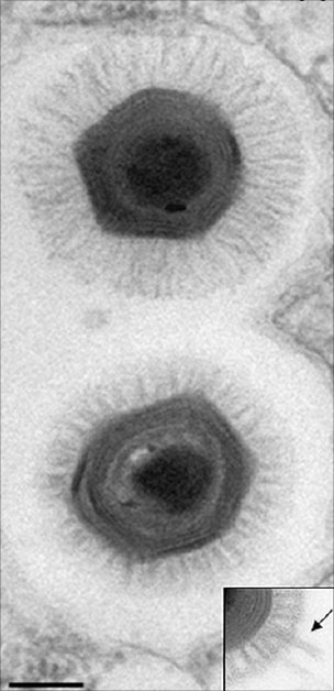
The team discovered that the HSV1 DNA is located very specifically in amyloid plaques: 90% of plaques in Alzheimer's disease sufferers' brains contain HSV1 DNA, and most of the viral DNA is located within amyloid plaques. The team had previously shown that HSV1 infection of nerve-type cells induces deposition of the main component, beta amyloid, of amyloid plaques. Together, these findings strongly implicate HSV1 as a major factor in the formation of amyloid deposits and plaques, abnormalities thought by many in the field to be major contributors to Alzheimer's disease.

The team had discovered much earlier that the virus is present in brains of many elderly people and that in those people with a specific genetic factor, there is a high risk of developing Alzheimer's disease.

The team's data strongly suggest that HSV1 has a major role in Alzheimer's disease and point to the usage of antiviral agents for treating the disease, and in fact in preliminary experiments they have shown that acyclovir reduces the amyloid deposition and reduces also certain other feature of the disease which they have found are caused by HSV1 infection.

Professor Itzhaki explains: "We suggest that HSV1 enters the brain in the elderly as their immune systems decline and then establishes a dormant infection from which it is repeatedly activated by events such as stress, immunosuppression, and various infections.

http://brainblogger.com/2010/12/15/alzheimer’s-disease-vaccine-on-the-horizon/

HSV1 is ubiquitous, identified in approximately 90% of adults. Normally, an infection with HSV1 occurs in infancy, but the virus remains lifelong in the peripheral nervous system in a latent, inactive state. HSV1 can be reactivated later in life by stress, immunosuppression, fever or ultraviolet light exposure; HSV1 is the virus that causes cold sores. Researchers postulate that if HSV1 reaches the brain, the virus could cause damage consistent with AD. Likewise, HSV1 is already identified as the cause of herpes simplex encephalitis, a rare but serious brain disorder, which leaves survivors with memory loss and a loss of cognitive function, just as AD does.

HSV1 does not cause AD on its own. There are likely host factors that alter the risks for developing AD. Interestingly, a genetic component — the type 4 allele of the apolipoprotein E gene, which normally transports lipids in the body and repairs tissue damage — confers a high risk of AD when associated with HSV1. (The same genetic component is an increased risk factor for cold sores.)

Two hallmarks of AD are the presence of amyloid plaques and neurofibrillary tangles in the brain. Beta-amyloid, the primary component of the plaques, accumulates in the presence of HSV1 infections. Further, 90% of plaques evaluated from AD brains contained HSV1, and 72% of the virus DNA was associated with plaques; in normal, aged brains, which contain amyloid plaques at a much lower frequency than AD brains, 80% of the plaques contained HSV1, but only 24% of the viral DNA was plaque-associated. In normal brains, it is likely that there is a lesser production or greater removal of beta-amyloid, so it is less likely that HSV1 would be able to interact destructively inside the brain. Basically, HSV1 infection likely induces changes in gene expression in the brain, through its inflammatory and oxidative processes, that are damaging to the brain.

The only current pharmacological therapies approved for AD are acetylcholinesterase inhibitors and N-methyl-D-aspartic acid receptor inhibitors, which demonstrate symptomatic improvement, but do not treat the underlying cause of AD. The proposed involvement of HSV1 in AD has led to the possibility of the first potentially disease-modifying treatments in AD. Antiviral agents, such as acyclovir and valacyclovir, may be beneficial in preventing disease progression in AD patients. These agents inhibit the synthesis of viral DNA, preventing its spread throughout the body and the damage it causes. (Acyclovir is also being evaluated in the treatment of multiple sclerosis, owing to another herpes virus implicated in the development of that disease.) Alternatively, a vaccine to prevent the reactivation of HSV1 and prevent AD altogether is being evaluated in human trials. Anti-viral agents are the first attempt to prevent the pathogenesis of AD, rather than just treat the symptoms, offering hope to millions of current and future AD sufferers and their families.


Chackerian B (2010). Virus-like particle based vaccines for Alzheimer disease. Human vaccines, 6 (11) PMID : 20864801

Chackerian B, Rangel M, Hunter Z, & Peabody DS (2006). Virus and virus-like particle-based immunogens for Alzheimer’s disease induce antibody responses against amyloid-beta without concomitant T cell responses. Vaccine, 24 (37-39), 6321-31 PMID : 16806604

Hill JM, Zhao Y, Clement C, Neumann DM, & Lukiw WJ (2009). HSV-1 infection of human brain cells induces miRNA-146a and Alzheimer-type inflammatory signaling. Neuroreport, 20 (16), 1500-5 PMID : 19801956

Itzhaki RF, & Wozniak MA (2006). Herpes simplex virus type 1, apolipoprotein E, and cholesterol: a dangerous liaison in Alzheimer’s disease and other disorders. Progress in lipid research, 45 (1), 73-90 PMID : 16406033

Lukiw WJ, Cui JG, Yuan LY, Bhattacharjee PS, Corkern M, Clement C, Kammerman EM, Ball MJ, Zhao Y, Sullivan PM, & Hill JM (2010). Acyclovir or Abeta42 peptides attenuate HSV-1-induced miRNA-146a levels in human primary brain cells. Neuroreport, 21 (14), 922-7 PMID : 20683212

Sabbagh, M., & Berk, C. (2010). Latrepirdine for Alzheimer’s disease: trials and tribulations Future Neurology, 5 (5), 645-651 DOI: 10.2217/fnl.10.53

Wozniak MA, Mee AP, & Itzhaki RF (2009). Herpes simplex virus type 1 DNA is located within Alzheimer’s disease amyloid plaques. The Journal of pathology, 217 (1), 131-8 PMID : 18973185

Wozniak, M., & Itzhaki, R. (2010). Antiviral agents in Alzheimer’s disease: hope for the future? Therapeutic Advances in Neurological Disorders, 3 (3), 141-152 DOI: 10.1177/1756285610370069
 
May 11, 2008
19,547
1,192
126
In my conquest of understanding the proliferation of diseases, i noticed something interesting. That cancer of the large intestines is relatively common while cancer of the small intestines is relatively rare.

When looking from the perspective of carrying bacteria in the large intestines, the small intestines are almost sterile with respect to the amounts of bacteria present in the small intestines. I found so far that cancer from the small intestines seems to start in nearby tissue and then spread out towards the small intestines. Afcourse there is always an exception. That exception seems to be Crohn's disease. Crohn's disease seems to be an auto immune system disease. From what i have read, this does not seem to be a genetic disorder. However, there has been research done that suggests that having a certain type of gene makes a person more susceptible to Crohn's disease but is not the primary cause. Crohn's disease also seems to be a family trait(duh :rolleyes: to myself : Genes.).

But i was thinking, the bacterial culture is also passed along from mother and father to child. I wonder if every isolated family have their own specific bacteria ? It makes sense. That some diseases are not just genetic (meaning in this special scenario more susceptible towards a certain disease) But that the bacterial culture also plays a role. If i think about it, i would expect diversification because of school and playgrounds would level out bacterial differences between young children. But maybe this is not entirely the case. Because when the children are home with their parents again, they are exposed again to the home environment (aka the family bacterial culture).

Now i mentioned that the small intestines are almost sterile meaning almost no bacteria. Maybe i am wrong, but Crohn's disease seems to start mostly at the ileum. The ileum is the last part of the small intestines and advances into the large intestines at the cecum. Here the appendix is also connected.

I read once in some journal although i do not know if this is true , that the appendix could be a bacterial reservoir.

Does Anybody have more interesting opinions or news ?
 
Last edited:
May 11, 2008
19,547
1,192
126
Forgot to mention this :
Celiac disease, i do not understand this disease. Sometimes it is invoked by a gluten rich diet caused by gliadin found in the gluten.


Perhaps there is another interesting factor. I do not know for sure and here is an idea :
Simply put, gluten is a combination of proteins found in wheat made from the endosperm of grains. Fungi are very common in wheat. In the past a lot of demon sightings and witch burning happened because of local wheat from local villages was invested with certain fungi. When consumed, the byproducts of these fungi would cause people to hallucinate and have violent spasms and pain cramps. Giving the illusion to be possessed while in reality to be poisoned. But hey these where mid evil times and the churches where fond of spreading lies.

I do wonder if a prolonged exposure to a certain type of fungi in the intestines could cause an immune response that would attack seemingly random tissues throughout the entire body. The fungi would not cause any symptoms. But if the fungi would have been consumed while being infected by a fungi virus, this would make a seemingly unrelated cause and effect.
The virus could spread unnoticed until it is too late. The immune system starts to attack every tissue where viral proteins are found. The key factor here would be that the viral proteins resemble the gliadin peptide found to be the cause of Celiac disease.
 
May 11, 2008
19,547
1,192
126
This is certainly an interesting view :

http://www.wired.com/medtech/health/news/2004/10/65252

Most of the cells in your body are not your own, nor are they even human. They are bacterial. From the invisible strands of fungi waiting to sprout between our toes, to the kilogram of bacterial matter in our guts, we are best viewed as walking "superorganisms," highly complex conglomerations of human cells, bacteria, fungi and viruses.

That's the view of scientists at Imperial College London who published a paper in Nature Biotechnology Oct. 6 describing how these microbes interact with the body. Understanding the workings of the superorganism, they say, is crucial to the development of personalized medicine and health care in the future because individuals can have very different responses to drugs, depending on their microbial fauna.

The scientists concentrated on bacteria. More than 500 different species of bacteria exist in our bodies, making up more than 100 trillion cells. Because our bodies are made of only some several trillion human cells, we are somewhat outnumbered by the aliens. It follows that most of the genes in our bodies are from bacteria, too.

Luckily for us, the bacteria are on the whole commensal, sharing our food but doing no real harm. (The word derives from the Latin meaning to share a table for dinner.) In fact, they are often beneficial: Our commensal bacteria protect us from potentially dangerous infections. They do this through close interaction with our immune systems.

"We have known for some time that many diseases are influenced by a variety of factors, including both genetics and environment, but the concept of this superorganism could have a huge impact on our understanding of disease processes," said Jeremy Nicholson, a professor of biological chemistry at Imperial College and leader of the study. He believes the approach could apply to research on insulin-resistance, heart disease, some cancers and perhaps even some neurological diseases.

Following the sequencing of the human genome, scientists quickly saw that the next step would be to show how human genes interact with environmental factors to influence the risk of developing disease, the aging process and drug action. But because environmental factors include the gene products of trillions of bacteria in the gut, they get very complex indeed. The information in the human genome itself, 3 billion base pairs long, does not help reduce the complexity.

"The human genome provides only scant information. The discovery of how microbes in the gut can influence the body's responses to disease means that we now need more research into this area," said Nicholson. "Understanding these interactions will extend human biology and medicine well beyond the human genome and help elucidate novel types of gene-environment interactions, with this knowledge ultimately leading to new approaches to the treatment of disease."

Nicholson's colleague, professor Ian Wilson from Astra Zeneca, believes the "human super-organism" concept "could have a huge impact on how we develop drugs, as individuals can have very different responses to drug metabolism and toxicity."

"The microbes can influence things such as the pH levels in the gut and the immune response, all of which can have effects on the effectiveness of drugs," Wilson said.

The Imperial College research demonstrates what many -- from X Files stalwarts to UFO fanatics -- have long claimed: We are not alone. Specifically, the human genome does not carry enough information on its own to determine key elements of our own biology.


Imagine what the environment for an effect might have if pollution is part of that environment.
New strains arise because of evolutionary survival. New strains the body may not be able to cope with. The balance distorted. The symbiotic structure unstable.
If you live wrongly, only seeking worldly pleasures, then you are destroying your own environment. And in the end destroying yourself or by means of karma, your offspring. Greed and selfishness is not a good thing.
 
Last edited:
May 11, 2008
19,547
1,192
126
This research seems to provide more proof that there is a link between the mind and the micro organisms in our gut.

http://medicalxpress.com/news/2011-08-probiotics-brain-functioning.html

(Medical Xpress) -- It was just last year that a certain company selling a special probiotic enhanced yogurt was ordered by a U.S. court to stop suggesting in its advertisements that it's product had health benefits that went beyond the norm. Now, new evidence by Javier Bravo and colleagues at University College Cork, suggests the company may have been on to something. In their paper, published in the Proceedings of the National Academy of Science, the team describes how mice given the prbiotic Lactobacillus rhamnosus, showed signs of being less anxious and depressed and even had lowered levels of stress hormones.

Building on recent research that suggests there may be more of a gut-mind link than scientists have realized (such as depression and anxiety linked to bowel problems) Bravo and his team decided to look into probiotics and their possible impact on mood. In their research, they focused on Lactobacillus rhamnosus, a probiotic bacterium normally found in the gut, and which is also commonly found in various kinds of yogurt and other types of dairy products.

To find out if ingesting L. rhamnosus did indeed have any impact beyond normal nutritional value, the team fed half of a group of mice a broth heavily laden with the bacterium for a period of time; the other half were given the same broth without the probiotic. Afterwards, the mice were tested to see if any discernible behavioral changes resulted.

Bravo et al found that the mice that had been given the probiotic demonstrated less anxious type behaviors, such as more of a willingness to traverse narrow walkways or to venture out into wide open spaces, activities that are known to cause stress in mice. They also found that the mice that had eaten the probiotic were less likely give in to the sensation of drowning when put in water, a sign that normally indicates depressive behavior. And finally, they found that the treated mice also had lower levels of stress hormones in their blood.

To find more proof of the connection, the team then severed the vagus nerve in the test mice and found the behaviors and hormone levels reverted back to their norms. The Vagus nerve transmits information from the gut and other organs to the brain, thus having it severed removes any means of communication between the two.

The team also found that the brain neurotransmitter gamma-aminobutyric acid (GABA) was also changed in the mice that had been fed the probiotic, with higher levels found in some areas of the brain associated with depression and lower levels in some areas associated with anxiety.

All in all the team feels confident that they’ve found a clear link between probiotics and mood and behavioral changes in mice; the next step of course will be to find out if the same is true for people.

More information: Ingestion of Lactobacillus strain regulates emotional behavior and central GABA receptor expression in a mouse via the vagus nerve, PNAS, Published online before print August 29, 2011, doi: 10.1073/pnas.1102999108

Abstract
There is increasing, but largely indirect, evidence pointing to an effect of commensal gut microbiota on the central nervous system (CNS). However, it is unknown whether lactic acid bacteria such as Lactobacillus rhamnosus could have a direct effect on neurotransmitter receptors in the CNS in normal, healthy animals. GABA is the main CNS inhibitory neurotransmitter and is significantly involved in regulating many physiological and psychological processes. Alterations in central GABA receptor expression are implicated in the pathogenesis of anxiety and depression, which are highly comorbid with functional bowel disorders. In this work, we show that chronic treatment with L. rhamnosus (JB-1) induced region-dependent alterations in GABAB1b mRNA in the brain with increases in cortical regions (cingulate and prelimbic) and concomitant reductions in expression in the hippocampus, amygdala, and locus coeruleus, in comparison with control-fed mice. In addition, L. rhamnosus (JB-1) reduced GABAAα2 mRNA expression in the prefrontal cortex and amygdala, but increased GABAAα2 in the hippocampus. Importantly, L. rhamnosus (JB-1) reduced stress-induced corticosterone and anxiety- and depression-related behavior. Moreover, the neurochemical and behavioral effects were not found in vagotomized mice, identifying the vagus as a major modulatory constitutive communication pathway between the bacteria exposed to the gut and the brain. Together, these findings highlight the important role of bacteria in the bidirectional communication of the gut–brain axis and suggest that certain organisms may prove to be useful therapeutic adjuncts in stress-related disorders such as anxiety and depression.
 
May 11, 2008
19,547
1,192
126
Researcher at the university of Michigan have found out how our little friends deal with uranium. The pili seem to play an important role.

http://www.physorg.com/news/2011-09-bacteria-immobilize-uranium.html


(PhysOrg.com) -- For several years, researchers have known that certain kinds of bacteria are able to "feed" off certain metals by either adding or removing electrons from their structure, but until now, haven’t really understood how they do it. Now, new research by Gemma Reguera and her team at Michigan State University have shown that the bacteria do so by means of protein nanowires, called pili, which are hair-like appendages with electrical conductivity. They have reported their findings in the Proceedings of the National Academy of Sciences.

The team specifically set out to find out how a specific type of bacterium known as a Geobacter, in this case, G. sulfurreducens, are able to clean up nuclear waste left behind by the cold war in such places as Colorado mines. They, like other researchers, believed that the bacteria were able to do its work through use of pili. In order to find out for sure, they had to induce the specimens to actually grow some in the lab, something that had stumped others before them. To force them, Reguera and her team subjected G. sulfurreducens, to much more harsh conditions than had been done before, presuming that the bacteria wouldn’t resort to using its pili unless pressed.

The tactic worked and the team was able to cause G. sulfurreducens to grow a mass of pili, which allowed them to study how they interacted with uranium. They found that the pili served as a buffer of sorts, protecting the cell structure of the bacterium as they also allowed for adding electrons to uranium ions which causes it to become more water soluble and thus safer to handle and clean up.

The pili grow to enormous lengths (though they are very then - only a few nanometers) relative to the bacteria that produce them, forming a conductive and protective barrier that allows the bacteria to thrive in truly hostile environments.

The study, part of ongoing research into so-named bioremediation; using organisms to remove unwanted substances from soil and water, adds to the growing body of knowledge that scientists hope will one day soon provide a means for dealing with a wide variety of environmental pollutants.

As for Reguera and her team, they hope their research eventually leads to getting away from using biological bugs to clean up toxic environments and more towards creating tiny little programmed robots that can mimic their actions but can be more easily manipulated into doing exactly what is needed in particular circumstances.

More about pili :

A pilus (Latin for 'hair'; plural : pili) is a hairlike appendage found on the surface of many bacteria.[1][2] The terms pilus and fimbria (Latin for 'thread' or 'fiber'; plural: fimbriae) can be used interchangeably, although some researchers reserve the term pilus for the appendage required for bacterial conjugation. All pili are primarily composed of oligomeric pilin proteins.

Pili connect a bacterium to another of its species, or to another bacterium of a different species, and build a bridge between the interior of the cells. This enables the transfer of plasmids between the bacteria. An exchanged plasmid can code for new functions, e.g., antibiotic resistance. The pilus is made up of the protein pilin.

Dozens of these structures can exist on the bacteria. Some bacterial viruses or bacteriophages attach to receptors on sex pili at the start of their reproductive cycle.

Pili are antigenic. They are also fragile and constantly replaced, sometimes with pili of different composition, resulting in altered antigenicity. Specific host responses to old pili structure are not effective on the new structure. Recombination genes of pili code for variable (V) and constant (C) regions of the pili (similar to immunoglobulin diversity).

http://en.wikipedia.org/wiki/Pilus
 
May 11, 2008
19,547
1,192
126
Epi genetics.

EDIT:
I almost forgot to write that although the research from physorg is about plants, it is not unique to plants.
/EDIT


A simplified explanation.
What i understand of it, is that humans (and possibly all mammals) do not posses enough genes where each gene separately encodes for a certain function. The solution is to use multiple genes at the same time and for a certain amount of time. The idea is that the environment also has control on the use of multiple genes and the time these genes are active.

Here is my opinion :
Remember quorum sensing of bacteria ?
Remember also that the bacteria on the human body are also part of the environment but also live in symbiosis with the cells in our body. And that we have more bacteria living inside us and on our skin then we have human cells. Remember also that all these bacteria together have more dna then humans do. I would not be surprised if the bacteria can also influence human gene expression by use of epigenetics.
For example if a human family lives on an isolated location. Then that human family will over time evolve differently depending on the environment when compared to other human families that live on other locations on the planet with different environments. I would think it is possible that each human family does not only have their own specific family genes, but also their own specific bacterial culture. The idea is that human dna does not change that quickly. But bacterial dna changes and evolves a lot faster( in matter of hours). If bacteria that live in symbiosis with humans can adapt faster to a changing environment, then these bacteria might also be able to adapt humans faster by use of epigenetics. Instead of letting the complex human dna evolve with all the issues that rise with it(diseases because of genetic disposition), we have epigenetics where the smaller microorganisms evolve and we adapt automagically. However, this works by the grace that the microorganisms do not evolve into something that will kill us. That can happen when the environment is seriously polluted. Cancer victims will rise for example. Miscarriages and stillbirth and birth defects will rise in number then as well.

I do think it is save to say we are the result of the symbiosis between the cells of our bodies and the bacteria that live in it and on it.


http://www.physorg.com/news/2011-09-genes-destiny-hidden-code-dna.html

A "hidden" code linked to the DNA of plants allows them to develop and pass down new biological traits far more rapidly than previously thought, according to the findings of a groundbreaking study by researchers at the Salk Institute for Biological Studies.

The study, published today in the journal Science, provides the first evidence that an organism's "epigenetic" code - an extra layer of biochemical instructions in DNA - can evolve more quickly than the genetic code and can strongly influence biological traits.

While the study was limited to a single plant species called Arabidopsis thaliana, the equivalent of the laboratory rat of the plant world, the findings hint that the traits of other organisms, including humans, might also be dramatically influenced by biological mechanisms that scientists are just beginning to understand.

"Our study shows that it's not all in the genes," said Joseph Ecker, a professor in Salk's Plant Molecular and Cellular Biology Laboratory, who led the research team. "We found that these plants have an epigenetic code that's more flexible and influential than we imagined. There is clearly a component of heritability that we don't fully understand. It's possible that we humans have a similarly active epigenetic mechanism that controls our biological characteristics and gets passed down to our children. "

With the advent of techniques for rapidly mapping the DNA of organisms, scientists have found that the genes stored in the four-letter DNA code don't always determine how an organism develops and responds to its environment. The more biologists map the genomes of various organisms (their entire genetic code), the more they are discovering discrepancies between what the genetic code dictates and how organisms actually look and function.

In fact, many of the major discoveries that led to these conclusions were based upon studies in plants. There are traits such as flower shape and fruit pigmentation in some plants that are under the control of this epigenetic code. Such traits, which defy the predictions of classical Mendelian genetics, are also found in mammals. In some strains of mice, for instance, a tendency for obesity can pass from generation to generation, but no difference between the genetic code of fat mice and thin mice explains this weight difference.

Scientists have even found that identical human twins exhibit different biological traits, despite their matching DNA sequences. They have theorized that such unexplained disparities could be the work of epigenetic variation.

"Since none of these patterns of variation and inheritance match what the genetic sequence says should happen, there is a clearly a component of the 'genetic' heritability that is missing," Ecker said.

Ecker and other scientists have traced these mysterious patterns to chemical markers that serve as a layer of genetic control on top of the DNA sequence. Just as genetic mutations can arise spontaneously and be inherited by subsequent generations, epigenetic mutations can emerge in individuals and spread into the broader population.

Although scientists have identified a number of epigenetic traits, very little was known about how often they arose spontaneously, how quickly they could spread through a population and how significant an influence they could have on biological development and function.


"Perception of the extent of epigenetic variation in plants from generation to generation varies widely within our scientific community," said Robert Schmitz, a post-doctoral research in Eckers' laboratory and the lead author on the paper. "We actually did the experiment, and found that overall there is very little change between each generation, but spontaneous epimutations do exist in populations and arise at a rate much higher than the DNA mutation rate, and at times they had a powerful influence over how certain genes were expressed."

In their study, the Salk researchers and collaborators at Scripps Research Institute mapped the epigenome of a population of Arabidopsis plants then observed how this biochemical landscape had changed after 30 generations. This mapping consisted of recording the state of all locations on the DNA molecule that could undergo a chemical modification known as methylation, a key epigenetic change that can alter how certain underlying genes are expressed. They then watched how methylation states of these sites evolved over the generations.

The plants were all clones of a single ancestor, so their DNA sequences were essentially identical across the generations. Thus any changes in how the plants expressed certain genetic traits were likely to be a result of spontaneous changes in their epigenetic code - variations in the methylation of the DNA sites- not the result of variations in the underlying DNA sequences.

"You couldn't do this kind of study in humans, because our DNA gets shuffled each generation," Ecker said. "Unlike people, some plants are easily cloned, so we can see the epigenetic signature without all the genetic noise."

The researchers discovered that as many as a few thousand methylation sites on the plants' DNA were altered each generation. Although this represents a small proportion of the potentially six million methylation sites estimated to exist on Arabidopsis DNA, it dwarfs the rate of spontaneous change seen at the DNA sequence level by about five orders of magnitude.

This suggests that the epigenetic code of plants - and other organisms, by extension - is far more fluid than their genetic code.

Even more surprising was the extent to which some of these changes turned genes on or off. A number of plant genes that underwent heritable changes in methylation also experienced substantial alterations in their expression - the process by which genes control cellular function through protein production.

This meant that not only was the epigenome of the plants morphing rapidly despite the absence of any strong environmental pressure, but that these changes could have a powerful influence on the plants' form and function.

Ecker said the results of the study provide some of the first evidence that the epigenetic code can be rewritten quickly and to dramatic effect. "This means that genes are not destiny," he said. "If we are anything like these plants, our epigenome may also undergo relatively rapid spontaneous change that could have a powerful influence on our biological traits."

Now that they have shown the extent to which spontaneous epigenetic mutations occur, the Salk researchers plan to unravel the biochemical mechanisms that allow these changes to arise and get passed from one generation to the next.

They also hope to explore how different environmental conditions, such as differences in temperature, might drive epigenetic change in the plants, or, conversely, whether epigenetic traits provide the plants with more flexibility in coping with environmental change.

"We think these epigenetic events might silence genes when they aren't needed, then turned them back on when external conditions warrant," Ecker said. "We won't know how important these epimutations are until we measure the effect on plant traits, and we're just now to the point where we can do these experiments. It's very exciting."


Here is a horizon documentary about epigenetics ( i know, i posted in this thread before) :

http://video.google.com/videoplay?docid=4942166965081178368


And a song, it has something...

SH3 - Letters From The Lost Day

http://www.youtube.com/watch?v=0BDu7Sa5GDE
 
Last edited:
May 11, 2008
19,547
1,192
126
http://medicalxpress.com/news/2011-09-gut-bacteria-immune-cells-friendly.html


Yahooo. Continued progress ?


Researchers find gut bacteria teaches immune cells to see them as friendly

(Medical Xpress) -- Most people know that the gut (human or otherwise) has bacteria in it that helps in the proper digestion of food. But how these bacteria manage to evade destruction by the immune system has been a mystery. Now, new research by a group working out of Washington University in St. Lois, as described in their paper published in the journal Nature, shows that such bacteria mange to survive by teaching T cells to see them as friends, rather than foes.

In the study, led by Chyi-Song Hsieh, the team first sought to discern whether the there was something going on in the development of T cells themselves that would account for them ignoring bacteria in the gut. To do this, they implanted some of the special T cell genes found only in the gut, into the bone marrow of a mouse that had been genetically modified to not produce T cells (which is where they normally come from). And though the T cells did grow, they didn’t have the same properties as the gut T cells and thus it was deduced that it wasn’t the environment in which they were spawned that led to them ignoring gut bacteria.

Next, the team looked at the mice they had just studied - one group had donated normal gut T cells genes, the other had genetically modified bone marrow and genes added from the first. The first group had gut bacteria, while the second did not. They discovered however, that when the two groups of mice were allowed to exist in the same cage, the mice with the modified bone marrow soon also had the special T cells that allowed the foreign bacteria to exist in its gut, clearly demonstrating that the bacteria in the normal mouse had somehow (after transferring via shared water and food dishes, etc.) trained the T cells in the guts of the modified mice (and changed them in the process) to ignore them. The question now, is how.

In an addendum to the research, the team also found that in studying mice with colitis, a condition generally associated with problems regarding helpful bacteria in the gut, there appeared to be problems in maintaining the regulatory T cells needed for proper digestion.

Now that researchers have a better understanding of which agent is responsible for allowing good bacteria to exist in the gut, new treatments might soon be on the way for those suffering from such gut ailments as colitis and Crohn's disease. They’ll also quite naturally, be trying to figure out how the bacteria trains gut T cells to abide them.

More information: Peripheral education of the immune system by colonic commensal microbiota, Nature (2011) doi:10.1038/nature10434

Abstract
The instruction of the immune system to be tolerant of self, thereby preventing autoimmunity, is facilitated by the education of T cells in a specialized organ, the thymus, in which self-reactive cells are either eliminated or differentiated into tolerogenic Foxp3+ regulatory T (Treg) cells1. However, it is unknown whether T cells are also educated to be tolerant of foreign antigens, such as those from commensal bacteria, to prevent immunopathology such as inflammatory bowel disease2, 3, 4. Here we show that encounter with commensal microbiota results in the peripheral generation of Treg cells rather than pathogenic effectors. We observed that colonic Treg cells used T-cell antigen receptors (TCRs) different from those used by Treg cells in other locations, implying an important role for local antigens in shaping the colonic Treg-cell population. Many of the local antigens seemed to be derived from commensal bacteria, on the basis of the in vitro reactivity of common colon Treg TCRs. These TCRs did not facilitate thymic Treg-cell development, implying that many colonic Treg cells arise instead by means of antigen-driven peripheral Treg-cell development. Further analysis of two of these TCRs by the creation of retroviral bone marrow chimaeras and a TCR transgenic line revealed that microbiota indigenous to our mouse colony was required for the generation of colonic Treg cells from otherwise naive T cells. If T cells expressing these TCRs fail to undergo Treg-cell development and instead become effector cells, they have the potential to induce colitis, as evidenced by adoptive transfer studies. These results suggest that the efficient peripheral generation of antigen-specific populations of Treg cells in response to an individual’s microbiota provides important post-thymic education of the immune system to foreign antigens, thereby providing tolerance to commensal microbiota.
 
May 11, 2008
19,547
1,192
126
More progress...

World class scientist Professor Willem M. de Vos will explain next Monday how the microbes that are closest to our hearts – gut microbes – could underpin a new way of thinking about human biology. As well as looking at our own genes, we can now include those of our microbes in studies of human health and disease. This is a significant shift in the way we approach human biology.


Gut microbes affect our health by producing vitamins, priming our immune system and contributing to resistance to pathogens. For example, recent studies have shown that the insulin resistance of patients with type 2 diabetes is linked to the intestinal microbiota composition and can be beneficially altered by replacing it with the microbiota of healthy donors.

The genes of our gut microbes, also known as the microbiome, act as a personalized organ that can be modified by diet, lifestyle and antibiotics. This organ is fed partly by us and partly by our diets. Professor de Vos and colleagues have classified the human microbiome into three enterotypes: clusters of microbiomes with similar compositions and nutrient-processing preferences. These enterotypes are characterized by bacteria with different capacities to degrade carbohydrate and mucin (a gel-forming protein which produces mucus). Our gut microbes get carbohydrates partly from our diet, whereas the mucin is produced by our own body.

Although these enterotypes are separated by species composition, it doesn't necessarily follow that abundant functions are provided by abundant species. To investigate the relationship between the microbiome and health, scientists must establish the functions of the products of their microbiomes.

"We have evolved with the microbes in our gut, our microbes inside, and have discovered that they talk to us and we feed them with, among other things, the mucins we produce. We now are trying to unravel their functions and understand exactly what these microbes and their products mean to human health" said Professor de Vos.

The size of one microbial metagenome (one host's microbiome) is 150 times larger than the human genome and encodes 100 times more genes than our own genome. This extensive gene catalogue could enable us to study potential associations between microbial genes and human phenotypes and even environmental factors like diet, throughout the length of our lifetime.

More information: On 10 October 2011, Professor Willem M. de Vos will present the fourth Environmental Microbiology Lecture: "Microbes Inside"


http://www.physorg.com/news/2011-10-gut-microbiome-human-health-disease.html
 
May 11, 2008
19,547
1,192
126
There is a virus discovered in 2010 that is huge. It is so big, that it can be seen with a normal visual light magnifying microscope. It is called Megavirus chilensis.

_55958338_55958337.jpg


http://www.bbc.co.uk/news/science-environment-15242386

The largest virus yet discovered has been isolated from ocean water pulled up off the coast of Chile.

Called Megavirus chilensis, it is 10 to 20 times wider than the average virus.

It just beats the previous record holder, Mimivirus, which was found in a water cooling tower in the UK in 1992.

Scientists tell the journal PNAS that Megavirus probably infects amoebas, single-celled organisms that are floating free in the sea.

The particle measures about 0.7 micrometres (thousandths of a millimetre) in diameter.

"It is bigger than some bacteria," explained Prof Jean-Michel Claverie, from Aix-Marseille University, Marseille, France.

"You don't need an electron microscope to see it; you can see it with an ordinary light microscope," he told BBC News.

Viruses cannot copy themselves; they need to invade a host cell if they want to replicate.

Like Mimivirus, Megavirus has hair-like structures, or fibrils, on the exterior of its shell, or capsid, that probably attract unsuspecting amoebas looking to prey on bacteria displaying similar features.

A study of the giant virus's DNA shows it to have more than a thousand genes, the biochemical instructions it uses to build the systems it requires to replicate once inside its host.

In the lab experiments conducted by Professor Claverie and colleagues, in which they infected fresh-water amoebas, Megavirus was seen to construct large trojan organelles - the "cells within cells" that would produce new viruses to infect other amoebas.

"Everything is initiated from a single particle, and then grows and grows to become this virion factory," explained Prof Claverie. "That's why it needs all these genes."

Megavirus was found off the coast of Las Cruces, central Chile. It was recovered as part of a general trawl in the ocean for biology of interest.

"This is a new way of doing virology," said Prof Claverie.

"Previously, we only discovered viruses because they caused disease in humans, or animals and plants. But now we are initiating what might be called environmental virology and we are looking for viruses everywhere.

"You just go to lakes, seas and oceans and pick up the water, and then you filter it, and try to rescue the virus by co-cultivating it with some potential host."

More generally, there is interest in ocean viruses because they have a major influence on populations of plankton, the microscopic organisms that form the base of many marine food chains. And when they kill plankton, viruses are also helping to regulate the planet's geochemical cycles as the dead organisms sink into the deep, locking away their carbon for aeons.

Prof Claverie said the megavirus would not be hazardous to humans.

http://en.wikipedia.org/wiki/Megavirus
 
May 11, 2008
19,547
1,192
126
There is new research that suggests that bacteria are exchanges genes at a faster rate then expected.

http://www.physorg.com/news/2011-10-bacteria-readily-swap-beneficial-genes.html

(PhysOrg.com) -- Much as people can exchange information instantaneously in the digital age, bacteria associated with humans and their livestock appear to freely and rapidly exchange genetic material related to human disease and antibiotic resistance through a mechanism called horizontal gene transfer (HGT).

In a paper appearing in Nature online Oct. 30, researchers — led by Eric Alm of MIT’s Department of Civil and Environmental Engineering and Department of Biological Engineering — say they’ve found evidence of a massive network of recent gene exchange connecting bacteria from around the world: 10,000 unique genes flowing via HGT among 2,235 bacterial genomes.

HGT is an ancient method for bacteria from different lineages to acquire and share useful genetic information they didn’t inherit from their parents. Scientists have long known about HGT and known that when a transferred gene confers a desirable trait, such as antibiotic resistance or pathogenicity, that gene may undergo positive selection and be passed on to a bacterium’s own progeny, sometimes to the detriment of humans. (For example, the proliferation of antibiotic-resistant strains of bacteria is a very real threat, as seen in the rise of so-called “superbugs.”)

But until now, scientists didn’t know just how much of this information was being exchanged, or how rapidly. The MIT team’s work illustrates the vast scale and rapid speed with which genes can proliferate across bacterial lineages.

“We are finding [completely] identical genes in bacteria that are as divergent from each other as a human is to a yeast,” says Alm, the Karl Van Tassel Associate Professor. “This shows that the transfer is recent; the gene hasn’t had time to mutate.”

“We were surprised to find that 60 percent of transfers among human-associated bacteria include a gene for antibiotic resistance,” adds computational systems biology graduate student Chris Smillie, one of the lead authors of the paper.

These resistance genes might be linked to the use of antibiotics in industrial agriculture: The researchers found 42 antibiotic-resistance genes that were shared between livestock-associated and human-associated bacteria, demonstrating a crucial link connecting pools of drug resistance in human and agricultural populations.

“Somehow, even though a billion years of genome evolution separate a bacterium living on a cow and a bacterium living on a human, both are accessing the same gene library,” Alm says. “It’s powerful circumstantial evidence that genes are being transferred between food animals and humans.”

Moreover, the team identified 43 independent cases of antibiotic-resistance genes crossing between nations. “This is a real international problem,” says microbiology graduate student Mark Smith, another lead author of the study. “Once a trait enters the human-associated gene pool, it spreads quickly without regard for national borders.”

The practice of adding prophylactic antibiotics to animal feed to promote growth and prevent the spread of disease in densely housed herds and flocks is widespread in the United States, but has been banned in many European countries. According to the Federal Drug Administration, more than 80 percent of the 33 million pounds of antibiotics sold in the United States in 2009 was for agricultural use, and 90 percent of that was administered subtherapeutically through food and water. This includes antibiotics such as penicillins and tetracyclines commonly used to treat human illness.

The MIT researchers found that HGT occurs more frequently among bacteria that occupy the same body site, share the same oxygen tolerance or have the same pathogenicity, leading them to conclude that ecology — or environmental niche — is more important than either lineage or geographical proximity in determining if a transferred gene will be incorporated into a bacterium’s DNA and passed on to its descendants.

“This gives us a rulebook for understanding the forces that govern gene exchange,” Alm says.

The team applied these rules to find genes associated with the ability to cause meningitis and other diseases, with the hope that transferred traits and the genes encoding those traits might make especially promising targets for future drug therapies.

“This is a very interesting piece of work that really shows how the increasing databases of complete genome sequences, together with detailed environmental information, can be used to discover large-scale evolutionary patterns,” says Rob Knight, associate professor of chemistry and biochemistry at the University of Colorado at Boulder. “The availability of vast datasets with excellent environmental characterization will give us an unprecedented view of microbes across the planet.”

Continuing the work, the researchers are now comparing rates of exchange among bacteria living in separate sites on the same person and among bacteria living on or in people with the same disease. They’re also studying an environmentally contaminated site to see which swapped genes might facilitate microbial cleanup by metal-reducing bacteria.

Other co-authors of the Nature paper are graduate student Jonathan Friedman, postdoc Otto Cordero and former graduate student Lawrence David, now at Harvard University.

Provided by Massachusetts Institute of Technology (news : web)
 
May 11, 2008
19,547
1,192
126
There is more research done in finding the causes of tumors in the colon.
The only strange from this research is that hydrogen peroxide causes a strong immune system response. Thus i personally think they got it backwards. I doubt that it is the immune response and the hydrogen peroxide that is the cause of colon tumors. I think it is more a response to the actual cause. There is more going on here. What these researchers claim here is that the immune response must be weakened. And that is wrong.


http://medicalxpress.com/news/2011-11-common-bacteria-colon-tumors-peroxide-producing.html

Working with lab cultures and mice, Johns Hopkins scientists have found that a strain of the common gut pathogen Bacteroides fragilis causes colon inflammation and increases activity of a gene called spermine oxidase (SMO) in the intestine. The effect is to expose the gut to hydrogen peroxide – the caustic, germ-fighting substance found in many medicine cabinets -- and cause DNA damage, contributing to the formation of colon tumors, say the scientists.

"Our data suggest that the SMO gene and its products may be one of the few good targets we have discovered for chemoprevention," says Robert Casero, Ph.D., professor of oncology at the Johns Hopkins Kimmel Cancer Center.

In a study, Casero and his colleagues introduced B. fragilis to two colon cell lines and measured SMO gene activity. In both cell lines, SMO gene activity increased two to four times higher than cells not exposed to the bacteria. The scientists also observed similar increases in enzymes produced by the SMO gene. The scientists successfully prevented DNA damage in these cells by blocking SMO enzyme activity with a compound called MDL 72527.

The Johns Hopkins team also tested their observations in a mouse model, created by Hopkins infectious disease specialist Cynthia Sears, M.D., to develop colon tumors. Mice exposed to the bacteria had similar increases in SMO. Mice treated with MDL 72527 had far fewer tumors and lower levels of colon inflammation than untreated mice. Results of the experiments were published online in the Proceedings of the National Academy of Sciences in August.

Casero says hydrogen peroxide can freely distribute through and into other cells. "It roams around, and can damage the DNA in cells," he says.

Rising levels of hydrogen peroxide and DNA damage in the colon are clear steps to tumor development, says Andrew Goodwin, Ph.D., who spearheaded the studies while performing graduate work in Johns Hopkins' Cellular and Molecular Medicine Program and Casero's laboratory.

B. fragilis strains that secrete a toxin are widely known to cause diarrhea in children and adults, and previous studies, including those at Johns Hopkins, have linked the toxin-producing bacteria to inflammation and colon cancer. Casero and collaborators previously linked the SMO gene to inflammation and cancer of the prostate and stomach.

Using MDL 72527 in humans is not advised, Casero says, because the compound blocks another enzyme in addition to SMO. Investigators hope to develop a drug that targets only the SMO enzyme. Candidates for such prevention strategies may include people with a history of colon polyps, which increases risk for colon cancer, and those with inflammatory bowel disease.


hydrogen peroxide and white blood cells.

http://news.sciencemag.org/sciencenow/2009/06/03-03.html

Anyone who has felt the sting as hydrogen peroxide foams and fizzes on a scraped knee knows about the compound's antiseptic properties. But new research suggests that hydrogen peroxide does more than just kill microbes. It may also call for reinforcements, summoning an army of bacteria-fighting cells to cuts and wounds.

Punctured skin sets off a chain reaction of chemical signals that activates blood-clotting and attracts an array of immune cells to guard against intruding microbes. Some of these cells, known as leukocytes, or white blood cells, kill by initiating a "respiratory burst," which releases highly reactive antimicrobial molecules, including hydrogen peroxide produced by the body itself.

Biologist Philipp Niethammer, a postdoctoral researcher at Harvard Medical School in Boston, was trying to coax such a hydrogen peroxide burst out of a nicked zebrafish tail when he noticed something odd. "I saw something bursting at the wound," he says, "but I didn't see leukocytes there." That bursting, experiments revealed, was hydrogen peroxide--appearing an average of 17 minutes before the arrival of the white blood cells that are supposed to produce it. To Niethammer, it appeared as if hydrogen peroxide was bringing leukocytes to the wound rather than the other way around.

To confirm the theory, Niethammer and his colleagues bathed zebrafish larvae in compounds known to inhibit the production of hydrogen peroxide. When researchers nicked larvae tails in the presence of the inhibitors, leukocytes stayed away: An average of fewer than one per larvae appeared at the cut within 42 minutes, compared with four to six under normal conditions. Next, the team used genetic manipulation to pinpoint the enzyme responsible for producing hydrogen peroxide. The culprit, a protein known as duox, is also found in the thyroid, digestive tract, and lungs of humans. Asthma and other disorders result from excessive inflammation in these tissues, so duox may play a role in those conditions, the researchers report tomorrow in Nature.

Paul Martin, a cell biologist at the University of Bristol in the United Kingdom, says the work identifies a key time point in wound healing. "Now we know the first step," he says. So, can that brown drugstore bottle of hydrogen peroxide also bring leukocytes to a wound? That's an open question, says Niethammer. He's now investigating whether white blood cells detect hydrogen peroxide directly or whether the compound is part of a longer signaling chain.

3 pictures of hydrogen peroxide in a wound and the migration of white blood cells towards the wound.

Sliced. A zebrafish larvae tail 3 minutes, 17 minutes, and 61 minutes (top to bottom) after being cut. Hydrogen peroxide (red) emanates from the wound, fading to yellow and green as it dissipates through tissue.
Credit: Philipp Niethammer
200960331-thumb-200xauto-2113.jpg
 
Last edited:
May 11, 2008
19,547
1,192
126
The mystery of resistance to the malaria parasite Plasmodium falciparum has been solved and explained. This is good news, because understanding these mechanisms will allow for better treatments against malaria and may be helpful in other diseases.

http://medicalxpress.com/news/2011-11-mystery-resistance-malaria.html

(Medical Xpress) -- Malaria is a disease caused by parasites passed to humans via the bites of infected mosquitoes. Globally, the disease causes over a million deaths every year, and is especially rife in parts of Africa and Asia. The parasites infect red blood corpuscles (the hemoglobin-containing cells that carry oxygen around the body) and hijack the support structure within the cells. Some people are known to be naturally resistant to the serious effects of malaria, and scientists have wondered for decades exactly how their resistance functions. Now new research gone a long way to solving the mystery.

It has been known for decades that some people in Africa and elsewhere who have a mutated gene that causes sickle cell anemia also have resistance to malaria because their red blood corpuscles contain an unusual form of hemoglobin―hemoglobin S, which results in the hemoglobin aggregating within the cell. Possessing only one copy of the mutated hemoglobin S makes the person a largely asymptomatic carrier, while two copies produces symptomatic sickle-cell anemia. In both cases the mutation gives some protection against malaria. Another mutation, hemoglobin C, causes hemolytic anemia when two copies of the mutation are present, and this form also protects against malaria.


plasmodiumfa.png


In a paper published in Science, researcher Marek Cyrklaff, of Heidelberg University in Germany, and colleagues in Germany and Burkina Faso, report that the unusual forms of hemoglobin in the red cells prevent the malaria parasite, Plasmodium falciparum, from hijacking the actin filaments that provide the skeleton scaffolding within the cell. They compared healthy and infected red corpuscles containing 'normal' hemoglobin with healthy and infected cells containing hemoglobin S or hemoglobin C. Using powerful cryoelectron tomography, the scientists discovered that in normal healthy cells the filaments of actin protein are short and located beneath the outer cell membrane , where they provide a support structure for the cell and makes it strong but pliable enough to pass through the tiniest blood vessels.

In infected cells with normal hemoglobin they found the actin protein was in long filaments, which the parasite used to build a cytoskeleton, or intracellular bridge, within the cell to transport its own manufactured protein, adhesin, to the surface of the cell. The effect of adhesin, as its name suggests, is to make adjoining cells stick together and to stick the cells to the blood vessel walls, causing the inflammation responses characteristic of malaria. In the hemoglobin S and C cells, the bridge could not be completed and the adhesin could not be effectively transported to the cell surface, thus reducing cell stickiness.

The scientists also found, after further experiments, that hemoglobin C and S are more easily oxidized than the unmutated form, and when actin filaments were placed with the hemoglobin, the C and S forms resulted in shorter actin filaments than normal hemoglobin, as did oxidized hemoglobin.

Malaria is most often treated with quinine, but clinical trials of a vaccine are now being carried out in Africa by GlaxoSmithKline, and the results look promising, with a 65% effectiveness rate. The new research suggests that further drugs could eventually be developed that interfere with the parasite's ability to use the actin filaments for its own purposes.

More information: Hemoglobins S and C Interfere with Actin Remodeling in Plasmodium falciparum–Infected Erythrocytes, Science, DOI: 10.1126/science.1213775

ABSTRACT
The hemoglobins S and C protect carriers from severe Plasmodium falciparum malaria. Here, we found that these hemoglobinopathies affected the trafficking system that directs parasite-encoded proteins to the surface of infected erythrocytes. Cryoelectron tomography revealed that the parasite generated a host-derived actin cytoskeleton within the cytoplasm of wild-type red cells that connected the Maurer's clefts with the host cell membrane and to which transport vesicles were attached. The actin cytoskeleton and the Maurer's clefts were aberrant in erythrocytes containing hemoglobin S or C. Hemoglobin oxidation products, enriched in hemoglobin S and C erythrocytes, inhibited actin polymerization in vitro and may account for the protective role in malaria.


About the parasite :
http://en.wikipedia.org/wiki/Plasmodium_falciparum
 

thaugen

Senior member
May 31, 2000
208
0
76
I'm not technically well-versed to enter the conversation. In fact I tried to write a short paper for lay persons on a different website with my understanding about how diet could prevent or help reverse cancer. It had to be short, not too technical, but hit all the major points. I'd appreciate any feedback to better develop it.


There are thousands of advocates of raw foods and eating healthy who say it may prevent cancer and it may be a helpful adjuvant when treating cancer, along with supplements and vitamins. Their evidence is not based on your beloved scientific studies/clinical trials but rather on case studies, individual outcomes where the persons survived after changing their diet. Like the original poster, they are still alive so they have no need to propose a mechanism of action, do lab tests, find appropriate clinical trial participants, or wait five years for the results to be published in a scholarly journal.

Those infected with scientism would now be frothing at the mouth and screaming "anecdotal, anecdotal, anecdotal!" I'm not a scientist, but I'll do some homework for you:

Inflammatory bowel disease: a model of chronic inflammation-induced cancer.
Chronic inflammation is a well-recognized risk factor for the development of human cancer. Inflammatory bowel disease (IBD), including ulcerative colitis and Crohn's disease, is a typical longstanding inflammatory disease of the colon with increased risk for the development of colorectal carcinoma. Several molecular events involved in chronic inflammatory process may contribute to multistage progression of human cancer development, including the overproduction of reactive oxygen and nitrogen species, overproduction/activation of key arachidonic acid metabolites and cytokines/growth factors, and immunity system dysfunction. Multiple animal models of IBD have been established, and in general, these models can be mainly categorized into chemically induced, genetically engineered (transgenic or gene knock-out), spontaneous, and adoptive transferring animal models.
http://www.ncbi.nlm.nih.gov/pubmed/19347299

From this I get that chronic inflammation is a bad thing and inflammatory bowel disease (IBD) is a bad thing and immune system dysfunction is a bad thing. So let's look at whether a cancer diet could affect any of these. Dannon yogurt advertising tells us that 70% of our immune system is located in the digestive tract. I found no scholarly article contesting that figure. So the theory is that a damaged digestive tract might also include immune system damage, and together they could lead to cancer. If a cancer diet could help mend the digestive tract and the immune system, that would be a good thing. I'm not saying, cure cancer, I'm just saying give the person a means to help influence a positive outcome.

BTW, the government's main cancer agency doesn't bother to tell you that a good portion of your immune system is located in the digestive tract:
What is biological therapy? http://www.cancer.gov/cancertopics/treatment/biologicaltherapy
Biological therapy (BYE-o-loj-ee-cal THER-ah-py) is a type of treatment that works with your immune system. It can help fight cancer or help control side effects (how your body reacts to the drugs you are taking) from other cancer treatments like chemotherapy.
What is the difference between biological therapy and chemotherapy?
Biological therapy and chemotherapy are both treatments that fight cancer. While they may seem alike, they work in different ways. Biological therapy helps your immune system fight cancer. Chemotherapy attacks the cancer cells directly.
How does biological therapy fight cancer?
Doctors are not sure how biological therapy helps your immune system fight cancer. But they think it may:
Stop or slow the growth of cancer cells.
Make it easier for your immune system to destroy, or get rid of, cancer cells.
Keep cancer from spreading to other parts of your body.
What is my immune system and how does it work?
Your immune system includes your spleen, lymph nodes, tonsils, bone marrow, and white blood cells. These all help protect you from getting infections and diseases. [spleen is the only part of the digestive tract mentioned]

Anyways, let's get back to the cancer diet theory. "Antioxidants are natural biochemical substances that protect living cells from harmful free radicals. Free radicals are unstable molecules formed as result of normal metabolic processes in the body. Viruses, bacteria, stress and pollution can also cause free radical production. When left unchecked, free radicals can cause our DNA to mutate, leading to cancer and other degenerative diseases.
The vitamins A, C and E, beta-carotene and oligomeric proanthocyanidins (OPC) obtained from wholesome foods are some examples of antioxidants.

Antioxidants and phytochemicals give fruits and vegetables their color, flavor and aroma. Phytochemicals, or plant chemicals, are compounds unique to each fruit, vegetable and herb. Phytochemicals protect plants from sunlight and also ensure their survival. Research has shown phytochemicals to possess enormous healing and disease-preventing properties because of their amazing ability to nourish and strengthen the immune system."

Colon cancer linked to bacteria: Bacterium Linked to Colorectal Cancer in Two Independent Studies http://www.cancer.gov/ncicancerbulletin/101811/page3

In the digestive tract resident microflora (good bacteria, yeasts, fungi, etc.) contain a number of components able to activate innate and adaptive immunity. Disruptions of the normal gastrointestinal microflora, such as overgrowth of harmful bacteria or yeasts like Candida, can damage the digestive system and possibly lead to not only harming the immune system but also enabling toxic products from those nasty bacteria and yeasts to enter the bloodstream. Furthermore, the immune system gets mobilized to try to stop these invaders and also to assist in dealing with the digestive system damage. The results: the immune system causes inflammation as a necessary part of the attack and repair processes and we have inflammatory bowel disease and the chronic inflammation that can facilitate cancer, including cancers located distant from the digestive system.

in the last 48 months, a growing body of research is underscoring a very significant relationship between gut microflora, systemic low-grade inflammation, metabolism, blood lipids and fat storage
http://www.gutpathogens.com/content/3/1/1

Dysregulation of the intestinal immune response to normal bacterial flora was suggested to play a crucial role in several inflammatory and autoimmune diseases.
http://www.sciencedirect.com/science/article/pii/S0165247804000379

Some of the damage resulting from small bowel bacterial overgrowth is produced by the action of bacterial proteases which degrade pancreatic and intestinal brush border enzymes causing pancreatic insufficiency, mucosal damage and malabsorption. In more severe cases the intestinal villi are blunted and broadened and mononuclear cells infiltrate the lamina propria. Increased fecal nitrogen leads to hypoalbuminemia. Bacterial consumption of cobalamin lowers blood levels of vitamin B12. Med Hypoth 1986, 20:125-132.

OK, here is where the oncologists and the cancer diet advocates part ways. As recently as 2003, the Journal of Clinical Oncology, discussing patients who use alternative therapies, stated:

"A widely disseminated literature on unorthodox treatments exists in print and on the Web for patients interested in seeking alternative therapies. There has been a two-decade-long movement toward more natural methods to treat a host of diseases, including cancer, and there is a significant degree of magical thinking about the role of the bowel in contributing to malignant disease ..."

Maybe that's why Spellman and other oncodocs are clinging to the outdated notion that most cancers have no relationship to dysfunction in the digestive system.

Here we need to bring in the famous "leaky gut syndrome." You might want to read up on the controversy surrounding it: http://en.wikipedia.org/wiki/Leaky_gut_syndrome but regardless, what you need to know is that scientific researchers now study it as "increased gastrointestinal permeability." Physicians may diagnose it as small intestine bacterial overgrowth (SIBO), or systemic candidiasis.

As of 2008 many doctors and researchers had accepted leaky gut as a fact:

The gut-brain barrier in major depression: intestinal mucosal dysfunction with an increased translocation of LPS from gram negative enterobacteria (leaky gut) plays a role in the inflammatory pathophysiology of depression.
There is now evidence that major depression is accompanied by an activation of the inflammatory response system and that pro-inflammatory [products of bad intestinal bacteria] may induce depressive symptoms.
The results show that intestinal mucosal dysfunction characterized by an increased translocation of gram-negative bacteria (leaky gut) plays a role in the inflammatory pathophysiology of depression. It is suggested that the increased translocation may mount an immune response and thus inflammatory response system activation in some patients with major depression ... It is suggested that patients with major depression should be checked for leaky gut by means of the IgM and IgA panel used in the present study and accordingly should be treated for leaky gut. http://www.ncbi.nlm.nih.gov/pubmed/18283240

So while you're getting cancer due to inflammatory response system activation (Spellman's chronic inflammation) you may also be getting majorly depressed due to the same bacteria problems.

C'mon, get to the point you say. OK, eating an anti-cancer diet and including probiotics (the good bacteria in yogurt) can sometimes clear up leaky gut syndrome all on its own. In the case of small intestine bacterial overgrowth (SIBO), the infection may be so bad as to need certain antibiotics along with the probiotics. But wait a minute you say, what about those cancers that are distant from the gut? A recent study of probiotics used to control acne found:

Recent studies have shown that orally consumed pre and probiotics can reduce systemic markers of inflammation and oxidative stress [49-51]. Since the local burden of lipid peroxidation in acne is high, such that it appears to place a great demand upon blood-derived antioxidants [52], the ability of oral probiotics to limit systemic oxidative stress [53] may be an important therapeutic pathway. Oral probiotics can regulate the release of inflammatory cytokines within the skin [54], and a specific reduction in interleukin-1 alpha (IL-1-α), noted under certain experimental conditions [55], would certainly be of potential benefit in acne. In line with observations of internal antibiotic use, it is also true that oral encapsulated probiotics have the potential to change the microbial community at sites far removed from the gastrointestinal tract [56].
http://www.ncbi.nlm.nih.gov/pmc/articles/PMC3038963/?tool=pubmed

That last sentence, "change the microbial community at sites far removed from the gastrointestinal tract" is important if you believe that several cancers are caused by microbes. Researchers have already proven that bacteria and viruses cause certain cancers. The jury is still out as to how many more cancers result from bacteria, viruses, fungi, parasites or other microbes. Notice also they mentioned cytikines, which Spellman referred to:

"Many cancers produce something called cytokines which shut down our immune system from recognizing them as different and acting on that."

Since probiotics can regulate the release of inflammatory cytokines, is it possible they could interfere with that cancer mechanism? Another mechanism they might affect is apoptosis (cancer cell death.) Ceramides are signaling molecules that alert cells to perform apostosis, programming a cell to die.

Recently, relatively simple sphingolipid metabolites, such as ceramide and sphingosine-1-phosphate, have been shown to be important mediators in the signaling cascades involved in apoptosis, proliferation, and stress responses. Specifically, researchers showed that the lactic acid bacteria Streptococcus thermophilus, a species found in most yogurts, can increase ceramide production.
A Textbook of Molecular Biotechnology by Ashok K. Chauhan, Ajit Varma

Phytosphingosine lipids inhibit micro-organisms and their second-messenger function, and are therefore considered part of the body's natural defense system, and have bacteria-killing properties. Not only does this enable Phytosphingosine to prevent acne from forming, but recent studies in France have also shown it to act as an anti-inflammatory at concentrations as low as 1%

So there you have it. Eat a good anti-cancer diet, take probiotics, and stay away from the stuff Spellman said causes cancer.
 

Gibsons

Lifer
Aug 14, 2001
12,530
35
91
I really don't have the time to sort this out, but.

You're overgeneralizing when it suits you, and nitpicking the other side when it suits you.

One specific thing I have to point out re the "digestive system" comprising the immune system. Lymphoid tissue is part of the digestive system. Your statement implying we're being misled is telling, and frankly, wrong.

I don't think there's anything wrong with your general conclusion and advice, but the construction of the argument just bugs me.
 
May 11, 2008
19,547
1,192
126
There is another breakthrough in research about how heliobacter pylori can survive in stomach acid. Now imagine, that when you have multiple pathogens, that while bacteria share genes through horizontal gene transfer, one of those harmful bacteria acquires for a short while the ability to survive stomach acid... And then ends up in the digestive system... It is a case of throwing the dice, but even throwing dice has a finite number of possibilities and thus shall occur sooner or later.

http://www.physorg.com/news/2011-12-ulcer-cancer-causing-bacterium.html


A research team led by scientists at the Chinese University of Hong Kong is releasing study results this week showing how a bacterium, Helicobacter pylori, that causes more than half of peptic ulcers worldwide and that has been implicated in stomach cancer has managed for eons to turn the acidic environment of the human gut into one in which it can thrive.


Writing in a Journal of Biological Chemistry "Paper of the Week," the scientists say the information they have obtained about the pathogen's clever employment of acid neutralizers may inform those who are designing new drugs to blunt H. pylori's effects across the globe.

H. pylori are the only bacteria known to thrive in the human stomach. It remains unclear how the pathogens are transmitted, although researchers suspect they could be spread through contaminated food or water. The damage the bacteria do to the mucous coating of the gut allows stomach acid to eat away at the sensitive organ lining, causing ulcers.

Although more than half of the world's population has the infection, for reasons still not quite understood most never develop ulcers. In fact, existing antibiotics can cure 80 to 90 percent of ulcers caused by the pathogen. However, H. pylori over the years have become increasingly resistant to antibiotics. Some experts have attributed that resistance to the fact that doctors are quick to prescribe antibiotics to kill it even when patients show no symptoms.

"There is a pressing need to develop new drugs and alternative strategies to fight against H. pylori infection before the prevalence of antibiotic resistance gets out of hand," says Ivan Fong, the lead author on the JBC paper and a graduate student at the Chinese University of Hong Kong whose research is focused on the biochemical makeup of protein complexes that assist in H. pylori's survival.

Ivan Fong, a graduate student at the Chinese University of Hong Kong, studies the biochemical makeup of protein complexes that assist in H. pylori's survival. Kam-Bo Wong is a professor at the institution and oversaw Fong's recent project. Credit: Chinese University of Hong Kong
It's the pathogen's ability to persist within the acid bath in the human stomach that has made it such a successful, albeit harmful, vector, says Fong. "The key is its use of an enzyme called urease to neutralize gastric acid," he explains.

H. pylori produce urease to spur the breakdown of urea, a naturally occurring chemical in the body, so that urea can release ammonia and make the gut an environment in which the pathogens can thrive. But, unlike most other enzymes, urease doesn't start doing its job immediately after being produced by the bacterium; instead, two nickel ions have to be delivered to it, and then the enzyme can mature, so to speak, and thus allow H. pylori to begin their damaging work.

"As the survival of H. pylori depends on active urease, this is a life-or-death issue for the pathogen to ensure nickel ions are delivered to the urease," says Kam-Bo Wong, a professor who oversaw the project at the institution.

It's not entirely clear how H. pylori make sure that urease can mature and then neutralize the surrounding acid. But Wong's team focused on four proteins that they suspect are helpers: UreE, UreF, UreG and UreH.

Using X-ray crystallography, "which essentially performs the function of a molecular microscope to visualize proteins with atomic resolution," Fong explains, the team took snapshots of UreF and UreH. What they saw was that UreH morphs the shape of UreF to enable UreF to recruit a third player, UreG, to form the UreF-UreH-UreG complex. In other words, the three proteins hook up to collectively deliver nickel ions to the right place on urease. Once the nickel ions are in place, they serve like a flint to ignite the breakdown of urea into ammonia, which then neutralizes the stomach acids.

"So, now we have a better understanding of how the machine can assemble itself, as if a skillful mechanic were there for the job, and deliver the nickel ions," says Fong.

Importantly, the team also discovered that disrupting the formation of the crafty UreF-UreH-UreG complex does, in fact, inhibit the synthesis of active urease. They hope that the information they've obtained about the molecular structures of UreF and UreH will help in the design of drugs that will essentially muck up the works of the molecular machine.

"As active urease is the key to survival of H. pylori, designing drugs that target this complex may well be a viable strategy to eradicate the pathogen," says Wong.

More information: The abstract for the paper, titled "Assembly of the preactivation complex for urease maturation in Helicobacter pylori: Crystal Structure of the UreF/UreH complex," is available at http://www.jbc.org … 830.abstract

Provided by American Society for Biochemistry and Molecular Biology
 
Last edited:

thaugen

Senior member
May 31, 2000
208
0
76
Gibsons: Thanks for the critique. I'll rethink some.

William: SIBO Small Intestine Bacterial Overgrowth can be several kinds of bacteria. Am I correct in assuming heliobacter pylori originating in the stomach will pass into the small intestine and could then share genes through horizontal gene transfer? Or would the small intestine bacteria be able to migrate back into the stomach, share genes and proliferate there?
 
May 11, 2008
19,547
1,192
126
Gibsons: Thanks for the critique. I'll rethink some.

William: SIBO Small Intestine Bacterial Overgrowth can be several kinds of bacteria. Am I correct in assuming heliobacter pylori originating in the stomach will pass into the small intestine and could then share genes through horizontal gene transfer? Or would the small intestine bacteria be able to migrate back into the stomach, share genes and proliferate there?

I must first honestly mention that i am not a specialist. What i know of it is that bacteria can share genes quickly. I do not know all methods of how bacteria share genes. I know one method which is called plasmids. If the special tricks of heliobacter pylori are part of such a plasmid, in the sense that it can be shared easily, perhaps another bacteria can copy this method. Maybe not as good as heliobacter pylori does, but good enough for a large group of bacteria to survive the stomach and to end up in the intestines. But to be honest, Gibsons has far more detailed knowledge about these subjects then i do.

My opinion :
Action is reaction. Microorganisms control the planet. And ancient multi cellular life proved very beneficial as vessels to move around quickly to escape from threats(volcanic activity and acids), and because of the numbers, redundancy increased survival. Mathematicians can show that last part easily for you if you are interested. After that, group conscience started to arise. Our ancestors. The way large groups of bacteria and cells communicate, self organization turned into a group conscience with specific complex behavior.
From a digital signal alike behavior with fixed and limited responses at the bacterial level to a more analog signal alike behavior that allowed for more complex interactions (quorum sensing). From a certain perspective one could say that this is the noise of life, the dice of god, the dice Einstein refused to accept.
We are surrounded with EM radiation. that is what powers life, from a certain perspective, one could say that this broadband em radiation that surrounds all around us is the clock signal for atoms to combine to molecules, for molecules to combine to organic compounds, to form life and to support.
Fourier figured it out...

As a sidenote, there is something that i am interested in. The mucous membranes in the mouth, would hormones be able to pass through these mucous membranes into the bloodstream ? We know that toxins such as alcohol can pass through mucous membranes in the bloodstream. We know ways exist to circumvent the digestive system to end up in the bloodstream directly. I know one method is the skin(for example medicine patches or nicotine patches and various chemical poisons), another method are mucous membranes. And the lungs can be used to pass toxic elements into the bloodstream. (I know my English is lousy)

Another sidenote :
This thread is intended for people in the biological field to think about an interesting possibility. Someone may read this thread, and may have a eureka moment. :) And it is for people who have a general interest in how nature really works.
 
Last edited:
May 11, 2008
19,547
1,192
126
This is interesting : The norovirus.
It seems according to current research that the norovirus ends up in the small intestines. This is normally a sterile environment meaning there are no bacteria here to be found in a healthy person.
This virus causes severe gastroenteritis. What i find interesting is how is the virus causes the illness. Is it a mimicry problem ? Or is the virus able to survive the stomach acid and do havoc in the small intestines cell lining ?

http://en.wikipedia.org/wiki/Norovirus
 

Mr. Pedantic

Diamond Member
Feb 14, 2010
5,039
0
76
This is interesting : The norovirus.
It seems according to current research that the norovirus ends up in the small intestines. This is normally a sterile environment meaning there are no bacteria here to be found in a healthy person.
This virus causes severe gastroenteritis. What i find interesting is how is the virus causes the illness. Is it a mimicry problem ? Or is the virus able to survive the stomach acid and do havoc in the small intestines cell lining ?

http://en.wikipedia.org/wiki/Norovirus
In medical terms 'sterile' is only taken to mean contamination with cellular organisms. Therefore, while the small intestine is generally taken to be sterile (which isn't actually completely true anyway) this does not mean that virus particles are not resident.

In terms of pathophysiology, I would assume that the virus causes gastroenteritis in a similar way that most viruses cause disease - viruses infect intestinal epithelial cells, they die, releasing cytoplasm (i.e. ions and proteins) into the lumen of the intestine, which causes water to follow through osmosis. The virus may also cause direct exposure of lymph and blood vessels to the lumen causing direct release of tissue fluid and blood into the lumen (though this will cause bloody diarrhoea).
 
May 11, 2008
19,547
1,192
126
In medical terms 'sterile' is only taken to mean contamination with cellular organisms. Therefore, while the small intestine is generally taken to be sterile (which isn't actually completely true anyway) this does not mean that virus particles are not resident.

In terms of pathophysiology, I would assume that the virus causes gastroenteritis in a similar way that most viruses cause disease - viruses infect intestinal epithelial cells, they die, releasing cytoplasm (i.e. ions and proteins) into the lumen of the intestine, which causes water to follow through osmosis. The virus may also cause direct exposure of lymph and blood vessels to the lumen causing direct release of tissue fluid and blood into the lumen (though this will cause bloody diarrhoea).

Thank you. :)
I agree that sterile intestines does not really mean petri dish sterile but more "no lethal pathogen" sterile...

But the real question is, does the virus travel through the bloodstream ending up in the cells of the small intestine ?
Or is the virus swallowed with food consumed, ending up in the stomach and then ending up in the small intestines ? Because then this would mean the virus can survive stomach acid. And that is important for often it is mentioned that the stomach acid will prevent any bacteria and virus from entering the intestines. We know this is not true, but we do not know full details in every specific case. The issue is that there are still a lot of people in the medical field who claim that nothing survives the stomach acid, thus claiming that cannot be a path for pathogens to end up in the small and specifically large intestines.
 

Mr. Pedantic

Diamond Member
Feb 14, 2010
5,039
0
76
Thank you. :)
I agree that sterile intestines does not really mean petri dish sterile but more "no lethal pathogen" sterile...

But the real question is, does the virus travel through the bloodstream ending up in the cells of the small intestine ?
Or is the virus swallowed with food consumed, ending up in the stomach and then ending up in the small intestines ? Because then this would mean the virus can survive stomach acid. And that is important for often it is mentioned that the stomach acid will prevent any bacteria and virus from entering the intestines. We know this is not true, but we do not know full details in every specific case. The issue is that there are still a lot of people in the medical field who claim that nothing survives the stomach acid, thus claiming that cannot be a path for pathogens to end up in the small and specifically large intestines.
As far as bacteria are concerned, as far as I know only H. pylori can replicate in the stomach. However, many species of bacteria (such as Clostridium difficile) can form spores, or other forms, that are resistant to, among other things, the conditions of the stomach (for example, C. difficile in its spore state is resistant to antibiotics, heat, alcohol, many commonly used sterilizing agents, oxygen, etc). These can pass through the stomach unharmed. I suspect that what you're mishearing is that people say that nothing else can survive in stomach acid - this is completely true, but it's misinterpreted to mean that the HCl will kill anything it comes into contact with, which is not true.

Many intestinal parasites work similarly as well, and that is how they are transmitted - the active zoites multiply in the intestine and cause disease, and any that pass into the large intestine form cysts or spores that prevent the organism from dying until it is ingested again.

It's obviously possible for viruses to be resistant to acidic conditions.
 
Last edited:
May 11, 2008
19,547
1,192
126
I really meant that some people in the biological field and medical have more then often claimed that nothing can pass the stomach acid. And they also mentioned that this is the case for viruses and bacteria, meaning these do not survive in any way at all. Of course this is a blatant lie and not deliberate at all, just uninformed. But they present this to common people who have no understanding at all with wrong information. Saliva in combination with stomach acid is the perfect defense it is often claimed and that is just not true.
 
May 11, 2008
19,547
1,192
126
http://www.physorg.com/news/2012-02-deadly-bird-parasite-evolves-exceptionally.html


Evolution of this specific bacteria has increased in speed since Mycoplasma gallisepticum (or at least the bacteria infecting the finches) lost certain genes. Genes that seem to protect it from... ? Bacteriophages...


A new study of a devastating bird disease that spread from poultry to house finches in the mid-1990s reveals that the bacteria responsible for the disease evolves at an exceptionally fast rate. What's more, the fast-evolving microbe has lost a key chunk of its genome since jumping to its new host, scientists were surprised to find. The missing portion contained the genes that made up the microbe's immune system, researchers report in the February 9th issue of PLoS Genetics.

When thousands of wild house finches started dropping dead from a mysterious eye infection in the Washington, DC, area in the winter of 1994, scientists were puzzled.
The birds had red, swollen, crusty eyes that left them unable to see or forage for food, until they eventually died from starvation or predation. Researchers soon identified the cause — a bacterium called Mycoplasma gallisepticum, a common cause of respiratory infections in turkeys and chickens that was previously known to infect only poultry.
By the time biologist Geoff Hill spotted his first sick bird in Auburn, Alabama, in 1995, the disease had spread through the eastern part of the continent, as far north as Quebec and as far south as Florida. "This was a devastating pandemic," Hill said.
Since its discovery, the epidemic has spread as far west as California, and is estimated to have wiped out hundreds of millions of birds. But scientists are still far from understanding how Mycoplasma gallisepticum gained the ability to spread to house finches — which diverged from chickens and turkeys some 80-90 million years ago — or what turned it into such a sweeping killer.

The red, swollen, crusty eyes in this house finch are the result of a highly-contagious infection caused by the bacterium Mycoplasma gallisepticum. Credit: Photo by Geoffrey E. Hill.
In a new study in the journal PLoS Genetics, researchers compared the genomes of a dozen strains of Mycoplasma gallisepticum sampled from infected house finches between 1994-2007, in the years following the initial outbreak. Using a technique called pyrosequencing, "we can measure evolution on very short time scales," said co-author Scott Edwards of Harvard University. Instead of studying the host switch years after it happened, the researchers are able to track it in real time. "We're catching the switch in the act," he added.
In both poultry and house finches, the microbe has been evolving at frightening speed, they report. "It's evolving anywhere from ten to 100 times faster than previous estimates for any other bacterium," said Harvard graduate student and first author Nigel Delaney.
But when the researchers compared the DNA sequences of the poultry strains with those sampled from house finches, they found something surprising — since making the switch, some parts of the parasite's genome have begun to break down.
Mycoplasma gallisepticum has a tiny genome to begin with, with less than 1000 genes, Delaney said. But rather than acquire new genes to help it outwit its new host, the parasite has gradually lost more than 50 genes — particularly those that make up the microbe's immune system.
Mycoplasmas are parasites, but they also have parasites of their own, including naturally occurring viruses called bacteriophages. "One of the main functions [of the genes that were lost] is to help guard Mycoplasma against the attacks of bacteriophages," Edwards explained.
"It was surprising to see a part of the genome that was assumed to be so important suddenly become unimportant," Delaney added.
It seems crazy, but "scientists have seen the same phenomenon before in HIV," said co-author Allen Rodrigo of the National Evolutionary Synthesis Center in Durham, North Carolina. When HIV infects a new host, it doesn't encounter the same threats, so it loses the specific immune defenses that protected it in its former host, Rodrigo explained. These original defenses may be expensive to maintain, Rodrigo said. Studies show that the HIV strains that lose them are able to reproduce more quickly and spread.

"It's similar to the 'use it or lose it' principle," Edwards added.

Mycoplasma gallisepticum can't be transmitted to humans. It can infect other backyard birds —including American goldfinches, purple finches, evening grosbeaks and pine grosbeaks — but none with the devastating consequences like those seen in house finches.
Researchers still don't know which genetic changes enabled the pathogen to reach epidemic proportions in house finches. But if the house finch strains have lost the genetic machinery that protected them in poultry, then reintroducing the parasites to the bacteriophages of their former hosts could be one way to control the disease, the scientists say.
Determining which specific bacteriophages those are, and whether the remnants of the Mycoplasma immune system still provide some protection against them, will take much more work.
"But this study shows that there's a third player that's important to understand this pandemic, which are the bacteriophages. Nobody's looked at that so far. This is the first observation that they seem to matter," Delaney said.
More information: Delaney, N., S. Balenger, et al. (2012). "Ultrafast evolution and loss of CRISPRs following host shift in a novel wildlife pathogen, Mycoplasma gallisepticum." PLoS Genetics. http://www.plosgen … pgen.1002511
 
Last edited: