William Gaatjes
Lifer
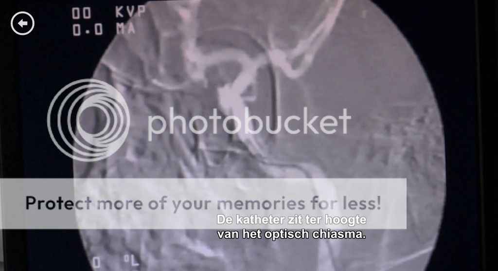
I am watching house and very often i see them guiding a tube like device through large aorta and large veins all the way inside the brain.
But they have this imaging system were they can see where they are inside the brain and it has a resolution high enough to actually see the veins.
Am i duped here and is this still science fiction or do such medical imaging systems actually exist ?
I know there exist a real time x ray imaging system that allows for to see the tissues while moving them around.
I had seen a youtube video about it once that explained how a woman had serious pain but was diagnosed as a fake because stationary imaging did not show any issues.
With that device i had seen on youtube, they could see that one of her vertebra in her neck slipped against the spinal cord causing her pain when she moved her head. This was something of a 30fps xray imaging system.
Or does House use a fluoroscope ?